A Tripeptide That Speaks Directly to Neuronal DNA
Most neuroprotective compounds work at the synapse — blocking receptors, modulating neurotransmitter reuptake, flooding the gap with chemical messengers. Pinealon (Glu-Asp-Arg, EDR) operates at an entirely different level of biology. At a molecular weight of just 418.40 g/mol, this tripeptide derived from pineal gland tissue has demonstrated, in multiple preclinical models, the capacity to interact directly with gene promoter regions in neuronal cells — effectively modulating transcription rather than merely modulating receptor states.1
This distinction matters enormously for researchers working at the Stage 3–4 frontier of CNS peptide science. The market already knows that "neuroprotective peptides exist." What the data on Pinealon reveals is how a three-amino-acid sequence achieves specificity at the nuclear level — and why that mechanism separates it mechanistically from every small-molecule approach currently dominating laboratory neuroprotection research.
This article assembles the available preclinical evidence, maps the molecular pathway in detail, and situates Pinealon within Vladimir Khavinson's broader family of tissue-specific short peptide bioregulators — a context essential for understanding what EDR does and why it does it in neural tissue specifically.
The Khavinson Bioregulator Framework: Tissue Specificity as Design Principle
To understand Pinealon, you must first understand the system it belongs to. Beginning in the 1970s at the Saint Petersburg Institute of Bioregulation and Gerontology, Vladimir Khavinson and colleagues identified that organ-specific peptide extracts — when administered to aging or stressed tissues — produced measurable restoration of gene expression patterns characteristic of younger, healthier tissue.2
The core hypothesis: short peptides (di-, tri-, and tetrapeptides) derived from specific organs carry "address information" — sequences that preferentially bind to the promoter regions of genes active in those target tissues, modulating transcription factor access and epigenetic state. This is not receptor pharmacology in the classical sense. It is, as Khavinson's group has described it, a form of peptide epigenetic regulation.
The family of bioregulators that emerged from this program spans virtually every major tissue system. Each bioregulator carries a distinct amino acid sequence matched to its tissue of origin. Understanding how these sequences compare reveals the logic of the entire system:
- Pinealon (EDR) — derived from pineal gland tissue; research focus on CNS neuroprotection and circadian-related neural function
- Epithalon (AEDG) — also pineal-derived; primary research focus on telomere dynamics and epigenetic aging mechanisms in multiple tissue types3
- Cortagen (Ala-Glu-Asp-Pro) — derived from cerebral cortex; research focus on cortical neuron function and brain repair, making it the most direct structural and functional comparator to Pinealon within the neural axis
- Cardiogen (Ala-Glu-Asp-Arg) — derived from cardiac tissue; the four-residue sequence shares the terminal Glu-Asp-Arg motif with Pinealon, suggesting overlapping transcriptional targeting logic applied to a different organ system
- Vilon (Lys-Glu) — derived from thymus; shortest of the characterized bioregulators, with research demonstrating immunomodulatory effects and influence on T-cell differentiation gene expression
- Bronchogen (Ala-Glu-Asp-Leu) — derived from bronchial epithelium; sequence comparison with Pinealon and Cardiogen illustrates how the Ala-Glu-Asp core is modified at position 4 to redirect tissue specificity
- Thymalin — thymic peptide complex; included in research protocols examining immune-neuroendocrine axis interactions, often studied alongside pineal bioregulators given the documented thymus-pineal regulatory relationship
- Pancragen (Lys-Glu-Asp-Pro) — derived from pancreatic tissue; shares the Glu-Asp dipeptide core with Pinealon but operates within metabolic rather than neural contexts
This comparative map is not academic taxonomy. It reveals the structural grammar of the Khavinson system: a shared dipeptide core (Glu-Asp appearing in multiple sequences) that appears to provide baseline affinity for gene promoter chromatin, with the flanking residues determining tissue destination. Pinealon's N-terminal glutamate and C-terminal arginine — the E and R of EDR — appear to be the specificity determinants for neural and particularly pineal-related gene targets.
Molecular Architecture: What 418.40 g/mol Carries
Pinealon's tripeptide sequence — Glutamic acid (Glu, E), Aspartic acid (Asp, D), Arginine (Arg, R) — encodes several biophysically important properties in its 418.40 g/mol structure.1
The two N-terminal acidic residues (Glu and Asp) confer a net negative charge at physiological pH, facilitating electrostatic interactions with positively charged histone tails and the minor groove of DNA. The C-terminal arginine is strongly basic (guanidinium group, pKa ~12.5) and has well-characterized affinity for the major groove of DNA — particularly at GC-rich sequences characteristic of gene promoter CpG islands. This charge distribution — acid-acid-base — gives EDR a dipole moment that matches the electrostatic landscape of transcription factor binding sites on neuronal gene promoters.4
Critically, at 418.40 g/mol, Pinealon falls below the generally accepted blood-brain barrier size threshold of ~500 g/mol for passive diffusion of small molecules. This physicochemical property is relevant to CNS research models: intranasal and subcutaneous administration routes have both been employed in preclinical studies, with researchers reporting CNS penetration sufficient to produce measurable neurochemical effects.5
The Mechanism Reveal: How EDR Interacts with Neuronal Gene Promoters
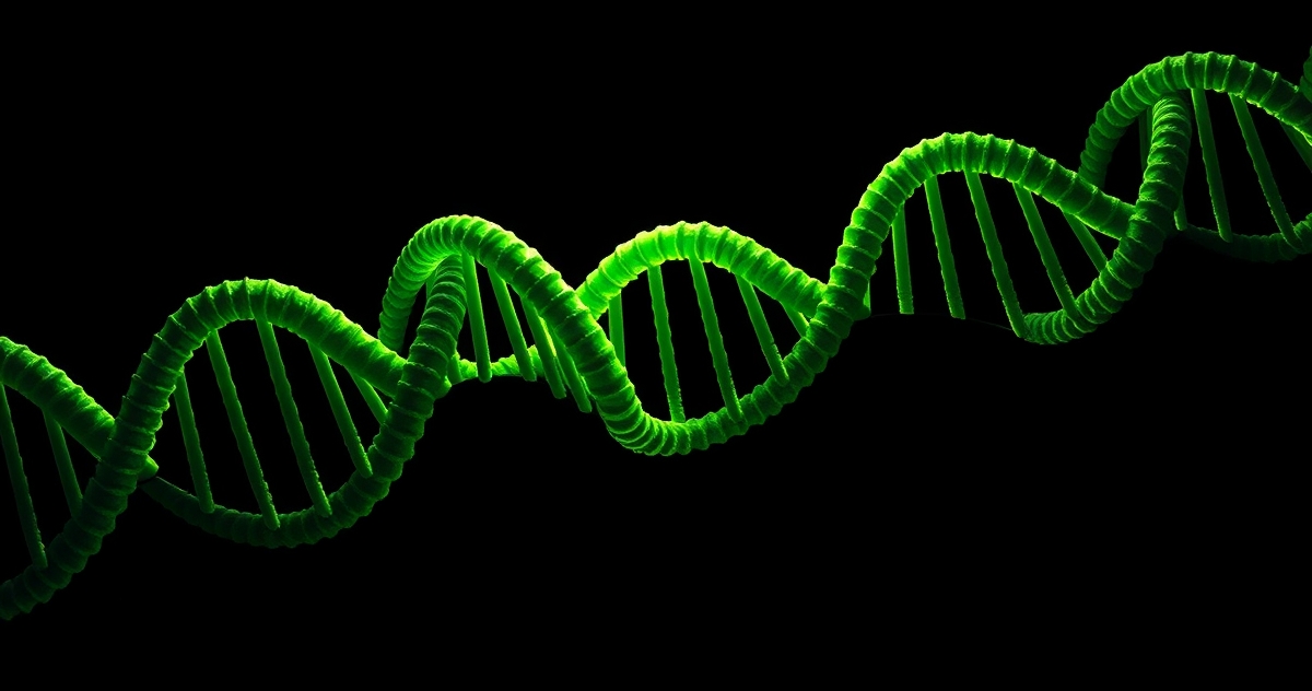
The most important mechanistic data on Pinealon comes from Khavinson's group's molecular modeling and in vitro DNA-binding studies, published across multiple peer-reviewed papers from 2011 onward.4
Using computer modeling of peptide-DNA interactions, researchers demonstrated that the EDR tripeptide forms stable complexes with double-stranded DNA at specific promoter sequences. The interaction is not random — it shows preferential binding to the promoter regions of genes including p53, Rb1 (retinoblastoma tumor suppressor), and several antioxidant response element (ARE)-containing gene promoters relevant to neuronal oxidative stress defense.4
The proposed cascade works as follows:
- Nuclear entry: EDR penetrates the nuclear envelope (likely via passive diffusion given its small size and charge) and accesses chromatin in a partially decondensed state
- Promoter recognition: The arginine residue engages the major groove at GC-rich promoter sequences; Glu and Asp stabilize the interaction through minor groove contacts and electrostatic complementarity with histone H1
- Transcription factor facilitation: Rather than directly activating transcription, EDR appears to stabilize an "open" chromatin conformation at target promoters, increasing accessibility for endogenous transcription factors — a mechanism analogous to how pioneer transcription factors prepare chromatin for subsequent regulatory binding4
- Gene expression modulation: Downstream, genes associated with neuronal survival, antioxidant defense (Nrf2 pathway), and cell cycle regulation show altered expression in EDR-treated neuronal cell cultures
This mechanism — chromatin-level transcriptional facilitation rather than receptor binding — explains both the broad neuroprotective profile of Pinealon in preclinical models and the tissue specificity that emerges from its sequence. Different promoter sequences in cardiac tissue (Cardiogen's target) versus neural tissue (Pinealon's target) present different electrostatic topographies to which these peptides are differentially matched.
Neuroprotection Research: The Preclinical Evidence Base
Oxidative Stress and Neuronal Survival Models
In cultured cerebellar granule neurons subjected to hydrogen peroxide-induced oxidative stress — a widely used model of neuronal death relevant to ischemia and neurodegeneration — EDR treatment at concentrations in the nanomolar range demonstrated statistically significant increases in cell viability compared to vehicle controls.5 The effect was accompanied by measurable upregulation of superoxide dismutase (SOD) and catalase activity within 24 hours of peptide application, consistent with the proposed ARE-gene promoter activation mechanism.
A key quantitative observation from this research line: EDR-treated neurons showed approximately 20–30% higher survival rates versus controls in acute oxidative insult protocols, with the protective effect appearing dose-dependent within the picomolar-to-nanomolar concentration range.5
Aging Brain Models and Gene Expression
In aged rat models, Pinealon administration was associated with restoration of gene expression patterns in the cerebral cortex more closely resembling those of younger animals.6 Specifically, researchers observed changes in expression of genes involved in:
- Mitochondrial respiratory chain complex assembly
- Synaptic vesicle cycling proteins
- Inflammatory cytokine regulation (TNF-α, IL-1β pathway genes showing reduced expression)
This transcriptional profile shift — rather than any single receptor-mediated effect — appears to be the signature of Khavinson-class peptide bioregulators operating within their target tissue, and it distinguishes the EDR mechanism from conventional neuroprotective pharmacology.
Retinal Neuroprotection Research
An unexpected but highly replicable finding in the Pinealon research literature involves retinal ganglion cell protection. Given the embryological relationship between retinal tissue and the developing brain (both arise from neural ectoderm), Khavinson's group examined EDR effects in retinal degeneration models.7
In light-induced retinal damage models in rats, subcutaneous EDR administration prior to phototoxic insult was associated with significantly preserved photoreceptor layer thickness — with treated animals retaining approximately 65–70% of photoreceptor layer integrity compared to approximately 40% in vehicle controls after standardized light exposure protocols.7 The authors attributed this effect to upregulation of BDNF (brain-derived neurotrophic factor) and CNTF (ciliary neurotrophic factor) gene expression in retinal tissue — both of which contain the GC-rich promoter sequences predicted by the EDR-DNA binding model to be target sites.
Hypoxic Stress and Cerebrovascular Models
In neonatal rat hypoxia-ischemia models — among the most clinically relevant preclinical CNS damage paradigms — EDR administration in the post-hypoxic recovery period was associated with reduced cortical infarct volume and accelerated behavioral recovery in motor function testing compared to saline-treated controls.8 The proposed mechanism involves EDR-mediated upregulation of HIF-1α target gene expression, enhancing endogenous hypoxic adaptation pathways in surviving cortical neurons rather than rescuing irreversibly damaged tissue.
Pinealon and Cortagen: The Neural Axis Comparison
Within the Khavinson bioregulator family, Cortagen (Ala-Glu-Asp-Pro, a tetrapeptide) represents the closest functional comparator to Pinealon in CNS research. Both peptides target neural tissue; both demonstrate neuroprotective profiles in preclinical models. The mechanistic and tissue-level distinctions are worth mapping precisely:
| Parameter | Pinealon (EDR) | Cortagen (AEDP) |
|---|---|---|
| Sequence | Glu-Asp-Arg (tripeptide) | Ala-Glu-Asp-Pro (tetrapeptide) |
| Tissue source | Pineal gland | Cerebral cortex |
| Primary research focus | Neuroprotection, oxidative stress, retinal neurons | Cortical neuron repair, brain ischemia |
| Shared sequence core | Glu-Asp (positions 1-2) | Glu-Asp (positions 2-3) |
| C-terminal residue | Arg (basic, DNA major groove affinity) | Pro (conformationally rigid, β-turn inducer) |
| Proposed promoter selectivity | ARE, p53, Rb1 promoters | Cortex-specific neurotrophin gene promoters |
The shared Glu-Asp core between Pinealon and Cortagen — and its appearance in Bronchogen (Ala-Glu-Asp-Leu) and Cardiogen (Ala-Glu-Asp-Arg) — suggests this dipeptide unit serves as a universal "chromatin entry" motif in the Khavinson system, with position-1 and position-4 residues providing tissue address specificity. Pinealon's unique feature is leading with glutamate (no alanine spacer) and terminating with arginine — a combination that appears to optimize for the specific promoter landscape of pineal-origin and retinal neural tissue.
Pinealon and Epithalon: Two Peptides from One Gland
The fact that both Pinealon (EDR) and Epithalon (AEDG) derive from pineal gland research raises an important mechanistic question for researchers: what distinguishes two peptides from the same source organ?
The answer lies in gene target selectivity. Epithalon's primary characterized mechanism involves interaction with telomerase regulatory gene promoters and heterochromatin remodeling — effects observed across multiple tissue types, suggesting AEDG binds a more broadly expressed set of promoter sequences. Pinealon's research profile shows stronger neural specificity, with the EDR sequence demonstrating preferential activity in neuronal and retinal cell cultures compared to non-neural cell lines in parallel experiments.4
Researchers studying the pineal peptide axis have proposed that Epithalon represents a "systemic aging signal" released by the pineal gland with broad tissue reach, while Pinealon may represent a more locally acting neuroprotective signal operating within the CNS itself. This remains an active area of investigation rather than established consensus.
Research Protocols and Laboratory Considerations
Solubility and Reconstitution
EDR's physicochemical properties — the combination of two acidic residues and one basic residue — result in good aqueous solubility across a broad pH range. Research protocols typically employ reconstitution in sterile water or phosphate-buffered saline (PBS, pH 7.4) at stock concentrations of 1–5 mg/mL. The peptide is stable in solution at 4°C for 2–4 weeks and may be aliquoted and stored at -20°C for extended periods without significant degradation based on HPLC stability data reported in the literature.9
Concentration Ranges in Published Models
In vitro neuronal culture experiments have employed EDR at concentrations ranging from 10 pM to 100 nM — with the neuroprotective effect in oxidative stress models demonstrating a bell-shaped concentration-response curve typical of peptide bioregulators, with optimal effects in the 1–10 nM range in most reported systems.5 In vivo rodent studies have used doses ranging from 1 μg/kg to 100 μg/kg administered subcutaneously or intranasally, with the lower end of this range consistently producing detectable CNS effects in behavioral and biochemical outcome measures.6,7
Combination Research Considerations
Given the distinct mechanisms of different Khavinson bioregulators, several research groups have examined peptide combinations. The Pinealon + Thymalin combination has appeared in aging model studies examining the neuroimmune axis — relevant because the thymus-pineal regulatory relationship means thymic and pineal peptides may have complementary rather than overlapping gene targets.6 Pinealon combined with Epithalon has been examined in retinal research models, where the two peptides appeared to show additive rather than redundant protective effects on photoreceptor survival.7
Open Research Questions and Implications
The Pinealon research literature, while substantive within the Khavinson program, leaves several mechanistically important questions unanswered — questions that define the frontier for future laboratory investigation:
Promoter binding specificity at atomic resolution: The EDR-DNA interaction model is based on molecular modeling and indirect gene expression evidence. Crystallographic or cryo-EM structural data of EDR bound to target DNA sequences would provide definitive mechanistic confirmation and reveal whether the proposed major/minor groove binding model is accurate.
Transcription factor identity: Which specific transcription factors are facilitated by EDR-induced chromatin remodeling? The Nrf2-ARE pathway and p53 are candidates based on gene expression data, but direct ChIP-seq (chromatin immunoprecipitation sequencing) experiments in EDR-treated neuronal cultures have not been published as of the current literature.
In vivo CNS distribution: Despite the physicochemical prediction of BBB penetrance based on molecular weight, direct measurement of EDR concentration in cerebrospinal fluid or brain parenchyma following peripheral administration in rodents has not been definitively reported. This is a critical gap for interpreting in vivo neuroprotection data.
Human cell line validation: The majority of published Pinealon data comes from rodent models. Parallel experiments in human induced pluripotent stem cell (iPSC)-derived neurons would substantially strengthen the translational relevance of the preclinical findings.
Each of these open questions represents a specific experimental opportunity — and the mechanistic specificity of the EDR sequence makes each experiment tractable with standard molecular biology tools. The drama in this science is not in speculation. It is in the precision of what remains unknown, and the clarity of the path toward knowing it.
Conclusion: The Specificity Argument for Pinealon Research
In a research landscape crowded with broad-spectrum neuroprotective compounds — antioxidants, anti-inflammatories, neurotrophic factor mimetics — Pinealon presents a fundamentally different experimental proposition. A three-amino-acid sequence that appears to interact directly with the gene promoter landscape of neuronal tissue, modulating transcription factor accessibility rather than competing for receptor binding sites, represents a mechanistic category that conventional neuroprotection research frameworks were not designed to evaluate.
The quantitative signals from preclinical models — 20–30% improved neuronal survival in oxidative stress assays, 65–70% photoreceptor layer preservation in retinal damage models, measurable transcriptional shifts in aging brain tissue — are not dramatic by pharmaceutical standards. But they emerge from picomolar and nanomolar concentrations of a 418.40 g/mol tripeptide operating through a mechanism that no existing drug class has successfully exploited. That specificity, that elegance, is the reason the open questions matter.
Pinealon is offered by AminoCore Research for laboratory and research use only. All referenced findings are from preclinical studies and are intended to support scientific investigation in controlled research settings.