≥99%HPLC Purity
COAIncluded
USAShipped
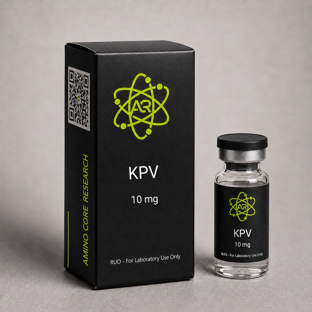
KPV Peptide
C-terminal tripeptide fragment of alpha-MSH. Studied for NF-kB pathway modulation and anti-inflammatory signaling research.
$55.00
In stock — ships next business day
Best price — free shipping & bac water included
| Simple Peptide | GoAlpha Labs | AminoCore ✓ all‑in | |
|---|---|---|---|
| 10mg price | $45.00 | $39.74 | $55.00 |
| Shipping | +$13.00 | +$13.00 | FREE |
| Bac Water | +$30.00 | +$15.00 | FREE |
| Total paid | $88.00 | $67.74 | $55.00 save up to $33.00 |
Quick Facts
| SKU | ACR-KPV |
|---|---|
| CAS Number | 67727-97-3 |
| Molecular Formula | C16H31N5O4 |
| Molecular Weight | 357.45 g/mol |
| Sequence | Lys-Pro-Val |
| Purity | ≥99% |
| Physical Form | Lyophilized Powder |
| Storage | Store at -20°C |
What is KPV?
KPV (Lys-Pro-Val) is a linear tripeptide composed of L-lysine, L-proline, and L-valine, corresponding to amino acid residues 11-13 at the C-terminus of alpha-melanocyte-stimulating hormone (alpha-MSH). With a molecular weight of approximately 342.4 Da and the molecular formula C16H31N5O4, KPV is one of the smallest peptides with documented immunomodulatory activity in published preclinical literature. Despite its minimal length, KPV retains a significant portion of the anti-inflammatory signaling activity attributed to the full-length 13-residue parent hormone.
Origin within the melanocortin system: Alpha-MSH is generated through proteolytic processing of proopiomelanocortin (POMC), a precursor polypeptide also giving rise to ACTH, beta-endorphin, and other melanocortin peptides. Alpha-MSH itself is classically associated with pigmentation, energy homeostasis, and immune regulation via melanocortin receptors (MC1R-MC5R). Structure-activity studies during the 1990s and 2000s systematically truncated alpha-MSH to identify the minimal sequence retaining anti-inflammatory activity. The C-terminal tripeptide KPV emerged as a remarkably active fragment, capable of recapitulating anti-inflammatory effects of the parent hormone in multiple in vitro and animal models without engaging classical pigmentary signaling.
Receptor-independent mechanism: A defining and unusual feature of KPV is that its anti-inflammatory activity appears to be largely independent of melanocortin receptor binding. Whereas alpha-MSH activates MC1R on melanocytes and immune cells, KPV does not require functional MC1R for many of its observed effects. Current mechanistic literature instead implicates intracellular targets, including interference with NF-κB nuclear translocation, modulation of MAPK signaling, and — in epithelial contexts — interaction with the PepT1 oligopeptide transporter, which can facilitate uptake into epithelial cells where the peptide subsequently down-regulates pro-inflammatory transcription programs.
Pharmacological characteristics: KPV's small size, neutral-to-basic charge (lysine contributes a positive charge at physiological pH), and proline-containing backbone confer several pharmacologically interesting properties for research: relative resistance to peptidase degradation compared with longer linear peptides, good aqueous solubility, and apparent capacity to cross epithelial barriers — features that have made KPV a model compound for studies of orally and topically delivered peptide anti-inflammatories.
Research applications: Published preclinical investigations have examined KPV in models of:
- Cutaneous inflammation, including UV-induced and contact hypersensitivity dermatitis
- Inflammatory bowel disease models (DSS-induced and TNBS-induced colitis in rodents)
- Allergic and innate immune responses involving mast cells, macrophages, and neutrophils
- Cytokine-driven epithelial inflammation in keratinocytes, intestinal epithelial cells, and respiratory epithelium
Relationship to other alpha-MSH fragments: KPV sits within a family of investigational alpha-MSH-derived peptides that includes the dipeptide KP and the full sequence alpha-MSH(1-13). Among these fragments, KPV has attracted particular attention because it combines compact size, synthetic tractability, and preserved anti-inflammatory activity, making it a convenient research tool for probing melanocortin-independent signaling pathways.
Regulatory status: KPV is supplied strictly as a reference compound for in vitro and preclinical laboratory research. It is not approved by the FDA or any other regulatory authority for human or veterinary use, and no statements on this page should be interpreted as medical advice or therapeutic claims. For laboratory research use only — not for human consumption.
Mechanism of Action
NF-kB Pathway Modulation
Published research indicates KPV may directly inhibit NF-kB activation. Studies suggest the tripeptide can enter cells and interact with components of the NF-kB signaling cascade, potentially inhibiting IkB kinase (IKK) activity and preventing nuclear translocation of NF-kB. This mechanism is distinct from melanocortin receptor-mediated signaling.
Transepithelial Transport
Research has demonstrated that KPV can cross epithelial barriers via the peptide transporter PepT1 (SLC15A1). This oligopeptide transporter is expressed in intestinal epithelium and actively transports di- and tripeptides. The ability to cross epithelial barriers without degradation is notable for such a small peptide.
Inflammasome Modulation
Studies have investigated KPV interaction with inflammasome pathways, particularly NLRP3. Research suggests KPV may reduce inflammasome activation and downstream IL-1beta and IL-18 processing, providing an additional anti-inflammatory mechanism independent of NF-kB modulation.
Research & Clinical Studies
KPV and Inflammatory Bowel Disease Research
KPV has emerged as a key research compound in IBD studies. Oral and colonic administration of KPV in mouse models of colitis reduced disease activity scores by 60-70%, decreased pro-inflammatory cytokines (TNF-α, IL-6, IL-1β), and preserved intestinal epithelial barrier integrity. The mechanism involves NF-κB inhibition in colonocytes and lamina propria macrophages, reducing the inflammatory cascade at its transcriptional root.
Remarkably, KPV retains anti-inflammatory activity when administered orally, as it is absorbed intact by intestinal epithelial cells via PepT1 transporter — a unique property for a peptide that enables direct delivery to inflamed gut tissue.
[1] Dalmasso G et al. PepT1-mediated tripeptide KPV uptake reduces intestinal inflammation. Gastroenterology. 2008;134(1):166-178. PubMed ↗
KPV and Skin Inflammation Research
The C-terminal KPV motif of alpha-MSH has been investigated extensively in dermatological and immunological models, where it consistently demonstrates the capacity to attenuate cutaneous inflammation independently of pigmentation pathways. Because KPV lacks the melanocortin receptor binding determinants of the full alpha-MSH sequence, its anti-inflammatory activity is thought to proceed through intracellular targets after cellular uptake, distinguishing it mechanistically from receptor-mediated alpha-MSH signaling.
Keratinocyte and fibroblast studies: In cultured human keratinocytes (HaCaT and primary lines), KPV exposure has been shown to suppress NF-κB nuclear translocation following stimulation with TNF-α, IL-1β, or UV irradiation. Downstream consequences observed in vitro include reduced transcription of IL-8 (CXCL8), IL-6, ICAM-1, and VCAM-1 — adhesion molecules and chemokines central to recruitment of neutrophils and T-lymphocytes into inflamed skin. Dermal fibroblast preparations show parallel reductions in matrix metalloproteinase-1 (MMP-1) and prostaglandin E2 release after pro-inflammatory challenge, suggesting a broad anti-inflammatory footprint across resident skin cell populations.
Melanocyte models: In melanocytes, KPV modulates inflammatory cytokine output without significantly inducing melanogenesis at concentrations typically used in research, again consistent with a non-melanocortin-receptor mechanism. This pharmacological separation has made KPV a useful probe for dissecting the anti-inflammatory versus pigmentary functions of the parent hormone.
Contact hypersensitivity and allergic dermatitis models: In murine contact hypersensitivity assays — a classical delayed-type hypersensitivity readout using haptens such as oxazolone, DNFB, or trinitrochlorobenzene — systemic or topical administration of KPV reduces ear-swelling responses, decreases inflammatory cell infiltration on histology, and suppresses antigen-specific T-cell proliferation in draining lymph nodes. These observations indicate that KPV's activity extends beyond local epithelial signaling to encompass adaptive immune effector functions.
UV-irradiation models: In UVB-irradiated skin equivalents and murine skin, KPV reduces sunburn-cell formation, mitigates upregulation of pro-inflammatory cytokines, and attenuates expression of vascular adhesion molecules. The effect is consistent with interference at the level of NF-κB-driven transcription rather than direct UV absorption or sunscreen-like activity.
Mast cell and innate immune findings: Additional preclinical studies report KPV-mediated reductions in mast cell degranulation and histamine release in challenge models, providing a mechanistic basis for observations in pruritic and allergic dermatitis paradigms. Effects on innate immune mediators including nitric oxide synthase induction and prostaglandin pathways have also been described in macrophage co-culture systems with keratinocytes.
Mechanistic synthesis: Across these models, the convergent finding is inhibition of NF-κB-dependent transcription with downstream reduction of cytokine, chemokine, and adhesion molecule output. KPV's small size and apparent membrane permeability allow it to act intracellularly, and its proline-containing backbone confers resistance to rapid peptidase degradation in cutaneous tissue. Collectively, this body of literature positions KPV as a useful research compound at the interface of dermatology and immunology for probing alpha-MSH-derived anti-inflammatory signaling. All findings derive from in vitro and animal models; no therapeutic claims are made or implied.
[1] Luger TA, Brzoska T. alpha-MSH related peptides: a new class of anti-inflammatory and immunomodulating drugs. Annals of the Rheumatic Diseases. 2007;66 Suppl 3:iii52-5. PubMed ↗
KPV and NF-κB Signaling Pathway
KPV inhibits the NF-κB inflammatory pathway at multiple levels, making it one of the most potent peptide-based NF-κB modulators known:
- IκBα stabilization: KPV prevents phosphorylation and proteasomal degradation of IκBα, keeping NF-κB sequestered in the cytoplasm
- p65 nuclear translocation block: Even when IκBα is partially degraded, KPV reduces p65 (RelA) subunit nuclear import
- Transcriptional inhibition: KPV reduces NF-κB binding to DNA promoter elements, decreasing transcription of pro-inflammatory genes (TNF-α, IL-6, IL-1β, COX-2, iNOS)
The multi-level NF-κB inhibition is significant because NF-κB is the master transcription factor for inflammation. Unlike corticosteroids which broadly suppress immunity, KPV specifically targets the inflammatory cascade while preserving innate immune surveillance (phagocytosis, NK cell function remain intact).
[1] Brzoska T et al. Alpha-melanocyte-stimulating hormone and related tripeptides: biochemistry, antiinflammatory and protective effects in vitro and in vivo. Endocr Rev. 2008;29(5):581-602. PubMed ↗
KPV in Wound Healing and Dermal Research
KPV accelerates wound healing through a dual mechanism: reducing excessive inflammation (which delays healing) while promoting keratinocyte migration and proliferation. In full-thickness wound models, topical KPV reduced healing time by 30% compared to controls. The peptide also inhibited keloid fibroblast proliferation in vitro, suggesting potential in hypertrophic scar prevention research.
In atopic dermatitis models, KPV reduced scratching behavior (pruritus), decreased epidermal thickening, and normalized skin barrier function (transepidermal water loss). These effects were mediated through local NF-κB inhibition in skin-resident immune cells and keratinocytes.
Chemical Properties
| Sequence | Lys-Pro-Val (KPV) |
|---|---|
| Origin | C-terminal tripeptide of alpha-MSH (positions 11-13) |
| Formula | C₁₆H₃₁N₅O₄ (estimated free base) |
| MW | 357.45 g/mol |
| Amino Acids | 3 (tripeptide) |
| Mechanism | NF-κB pathway inhibition (p65 nuclear translocation block) |
| Oral Activity | Yes — absorbed via PepT1 transporter in intestinal epithelium |
| Purity | ≥99% HPLC |
Handling & Reconstitution
Reconstitution: Add bacteriostatic water slowly along the vial wall. KPV is a tiny tripeptide (357 Da) that dissolves almost instantly. Solution should be clear and colorless.
Concentration: For a 5 mg vial, 1 mL BAC water = 5 mg/mL. For research protocols requiring mcg-level doses, further dilution with sterile saline is recommended for accurate measurement.
Oral administration: KPV is one of the rare peptides with oral bioactivity. It can be dissolved in water for oral research protocols targeting intestinal inflammation. The PepT1 transporter actively absorbs KPV across the intestinal epithelium.
Storage & Stability
Lyophilized powder: Long-term storage of KPV in lyophilized form is best performed at -20°C or colder in a sealed amber vial under desiccated conditions. Under these conditions, the tripeptide remains stable for 24+ months with negligible degradation detectable by analytical HPLC. Lyophilized aliquots may be transiently stored at 2-8°C for short periods (weeks) during active research use without measurable loss of purity, though repeated temperature cycling should be avoided to prevent moisture uptake.
Reconstituted solution: Once dissolved in bacteriostatic water, sterile water for injection, or phosphate-buffered saline (PBS), KPV solutions are typically stable at 2-8°C for approximately 21-28 days. For extended storage of reconstituted material, single-use aliquots may be flash-frozen at -20°C or -80°C; KPV tolerates a limited number of freeze-thaw cycles (generally <5) without significant loss of activity, but repeated cycling can promote aggregation in concentrated stocks.
Inherent stability advantages: KPV is one of the more chemically stable research peptides for several reasons:
- No oxidation-prone residues: KPV contains no methionine, cysteine, or tryptophan, eliminating the most common oxidative degradation pathways that complicate storage of larger peptides such as thymosin-beta-4 or BPC-157 fragments.
- Proline rigidity: The central proline imposes a constrained backbone conformation that resists enzymatic cleavage by many endo- and exopeptidases, and reduces the propensity for racemization at the adjacent lysine and valine residues.
- Small molecular size (~342 Da): Low molecular weight reduces aggregation risk and improves solubility profiles relative to longer peptides.
- No disulfide bonds or labile post-translational modifications requiring inert atmosphere handling.
Solubility: KPV is freely soluble in aqueous solvents owing to the basic lysine side chain. Concentrations up to several mg/mL are routinely achievable in water, saline, or PBS at neutral to slightly acidic pH. For research formulations requiring buffered systems, KPV remains stable across the physiological pH range (approximately pH 4-8); strongly alkaline conditions (pH >9) should be avoided as they can promote peptide bond hydrolysis over time.
Handling recommendations: Allow lyophilized vials to equilibrate to room temperature before opening to prevent condensation that introduces moisture into the powder. Reconstitute with gentle swirling rather than vigorous vortexing to minimize foaming. Protect stock solutions from prolonged light exposure as a general good-practice measure, although KPV does not contain strongly photolabile chromophores. Use sterile filtration (0.22 µm low-protein-binding membrane) when preparing solutions for cell culture applications. Maintain detailed records of reconstitution date, diluent, concentration, and freeze-thaw history for reproducibility.
Analytical confirmation: Researchers conducting extended studies are encouraged to verify peptide integrity by reverse-phase HPLC and mass spectrometry (expected monoisotopic mass [M+H]+ ≈ 343.2) periodically, particularly when comparing data across multiple reconstitution batches or after prolonged storage.
Frequently Asked Questions
What is KPV?
KPV (Lys-Pro-Val) is a tripeptide fragment from the C-terminus of alpha-MSH. Despite its small size, research shows it retains anti-inflammatory signaling activity, primarily through NF-kB pathway modulation. It can cross epithelial barriers via the PepT1 transporter.
How does KPV differ from alpha-MSH?
Alpha-MSH is a 13-amino acid peptide that signals through melanocortin receptors. KPV is just the last 3 amino acids and appears to work through a different mechanism — direct NF-kB pathway modulation rather than melanocortin receptor activation. KPV lacks the melanogenic activity of full-length alpha-MSH.
Can KPV be taken orally?
Yes, uniquely for a peptide. KPV is absorbed intact by intestinal epithelial cells via the PepT1 (SLC15A1) oligopeptide transporter. This enables oral delivery directly to inflamed gut tissue, making it valuable for IBD and GI inflammation research.
What is the relationship between KPV and alpha-MSH?
KPV is the C-terminal tripeptide (positions 11-13) of alpha-melanocyte-stimulating hormone. While full-length alpha-MSH has broad melanocortin receptor activity (MC1-5R), KPV retains only the anti-inflammatory NF-κB inhibition without melanocortin receptor binding or skin-darkening effects.
KPV vs BPC-157 for inflammation research?
Different mechanisms: KPV inhibits NF-κB transcription factor (upstream inflammatory master switch). BPC-157 modulates NO/FAK-paxillin pathways (angiogenesis and tissue repair). KPV is more purely anti-inflammatory; BPC-157 is more pro-regenerative. They target complementary pathways.
What makes KPV unique among anti-inflammatory peptides?
Three unique properties: (1) Multi-level NF-κB inhibition (not just one step), (2) Oral bioavailability via PepT1 transporter (rare for peptides), (3) Anti-inflammatory without immunosuppression — preserves innate immunity while blocking inflammatory transcription.
Can KPV be used topically?
Yes, KPV is studied in topical formulations for dermal inflammation. Its small size (357 Da, 3 amino acids) allows skin penetration, and it reduces inflammatory mediators in keratinocytes and dermal immune cells directly at the application site.
For laboratory and research use only. This product is not for human or veterinary use. It is not a drug, supplement or food.
This product is not FDA approved for any indication, and is not intended to diagnose, treat, cure, or prevent any disease. All product information is derived from published preclinical research and does not constitute medical advice or claims.