Introduction: A Central Mechanism in Melanocortin Research
Most compounds investigated for neuroendocrine signaling operate at the periphery — acting on blood vessels, smooth muscle, or tissue-level receptors far from the circuits that actually generate behavioral outputs. PT-141 (Bremelanotide) does something fundamentally different. It bypasses peripheral vasculature entirely and targets the melanocortin receptor system within the central nervous system itself, engaging MC3R and MC4R subtypes in hypothalamic nuclei that sit at the intersection of dopaminergic signaling, energy homeostasis, and motivational circuitry.1
This architectural distinction — central versus peripheral — is not merely pharmacological trivia. It defines what this compound can and cannot do in research models, what questions it can answer, and why it has attracted sustained attention from neuroscientists investigating the melanocortin system. Understanding PT-141 means understanding the receptor subtypes it targets, the cascade those receptors initiate, and the broader neuroendocrine network those cascades are embedded within.
This article assembles that picture in full — from the cyclic heptapeptide structure that makes selective CNS receptor engagement possible, through the hypothalamic dopaminergic pathway it modulates, to the research protocols that have used it to interrogate melanocortin system function. The market for peptide research has reached a stage where simply naming a compound and claiming it "activates receptors" is insufficient. What investigators require — and what the evidence supports — is the specific molecular story.
Structural Derivation: From Melanotan II to Bremelanotide
PT-141's lineage begins with alpha-melanocyte-stimulating hormone (α-MSH), the endogenous thirteen-residue peptide cleaved from proopiomelanocortin (POMC) that serves as the natural ligand for the melanocortin receptor family. α-MSH binds across MC1R through MC4R with varying affinity, producing the full range of melanocortin biological effects — pigmentation, energy balance, inflammation modulation, and neuroendocrine signaling.2
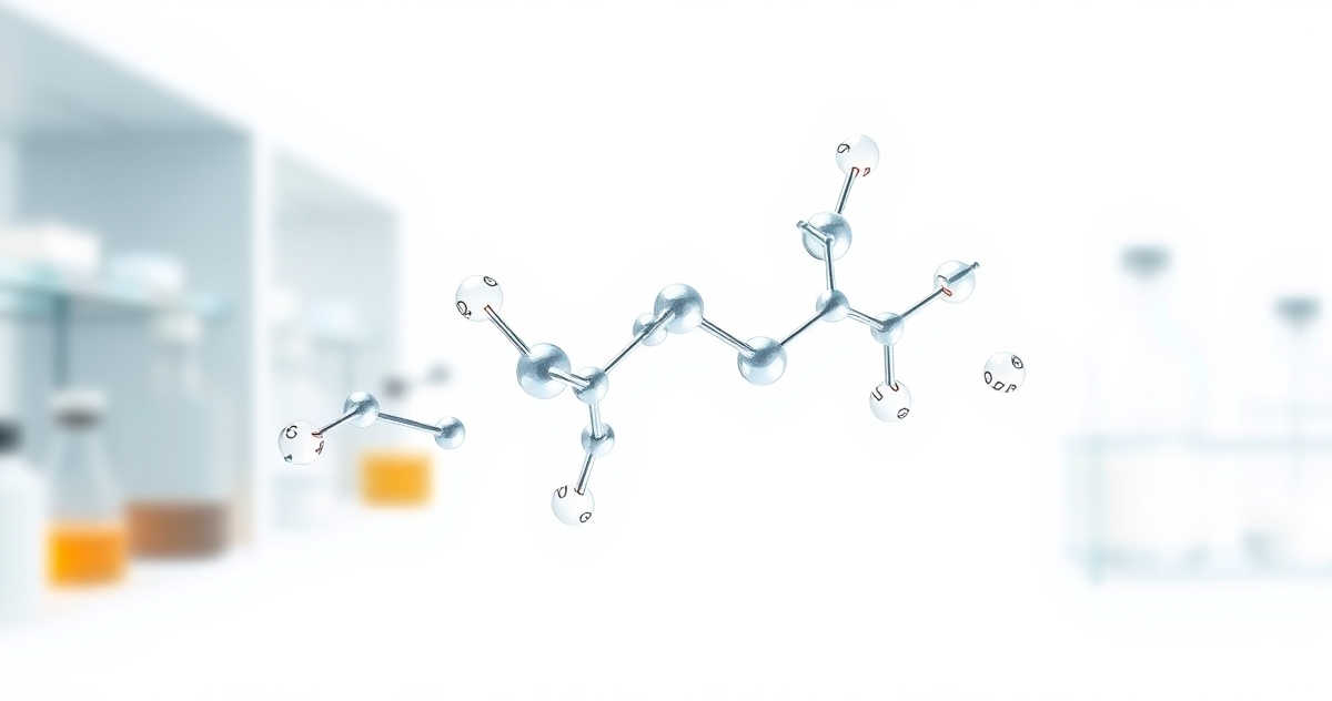
The first synthetic step toward PT-141 was Melanotan I (afamelanotide), a linear analogue of α-MSH with extended half-life engineered through substitution of a serine residue with norvaline and replacement of the methionine at position 4 with phenylalanine. Melanotan I showed improved stability but broad receptor engagement. The decisive structural advance came with Melanotan II, a cyclic lactam heptapeptide — Ac-Nle-cyclo[Asp-His-D-Phe-Arg-Trp-Lys]-NH₂ — that achieved dramatically increased potency through the conformational rigidity imposed by the cyclic structure.3
The cyclic lactam bridge between Asp⁴ and Lys¹⁰ locks the peptide backbone into the bioactive conformation required for high-affinity melanocortin receptor engagement. This constraint accomplishes two things simultaneously: it reduces the entropic cost of receptor binding (the peptide does not need to adopt the correct conformation upon binding — it already exists in that conformation), and it substantially increases resistance to peptidase degradation compared to linear analogues. The result is a compound with nanomolar affinity at MC3R and MC4R and a plasma half-life of approximately 60 minutes, compared to minutes for native α-MSH.3
PT-141 (Bremelanotide) is the metabolite of Melanotan II generated by removal of the acetylamide group. It retains the cyclic heptapeptide core — the same Nle-cyclo[Asp-His-D-Phe-Arg-Trp-Lys] scaffold — and the same receptor selectivity profile, with high affinity for MC3R and MC4R and lower affinity for MC1R and MC5R. The D-phenylalanine substitution at position 7 (replacing the native L-configuration) is critical: it is the primary driver of both increased receptor potency and CNS penetration relative to the parent α-MSH sequence.1
The Melanocortin Receptor Family: Subtype Architecture and Research Significance
Five melanocortin receptor subtypes (MC1R–MC5R) have been characterized, each encoded by distinct genes, expressed in distinct tissue distributions, and coupled to distinct downstream signaling programs. All five are class A G-protein-coupled receptors (GPCRs) that signal primarily through Gs-mediated adenylyl cyclase activation and consequent cAMP accumulation, though MC3R and MC4R also demonstrate coupling to additional G-protein subtypes in specific cellular contexts.4
For PT-141 research, the relevant subtypes are MC3R and MC4R. Their distribution and function define the biological questions this compound can investigate:
MC3R: Hypothalamic Energy Sensing and Dopaminergic Interface
MC3R expression is concentrated in the hypothalamus — particularly in the arcuate nucleus, ventromedial nucleus, and lateral hypothalamic area — as well as in the limbic system and brainstem. Critically, MC3R is expressed on dopaminergic neurons in the ventral tegmental area (VTA) and mesolimbic projections, positioning it as a direct modulator of dopaminergic neurotransmission.5 Research in MC3R knockout models has demonstrated disrupted energy balance, altered circadian feeding rhythms, and impaired dopaminergic tone in reward circuits, establishing MC3R as a neuromodulatory node connecting melanocortin signaling to motivational circuitry.5
MC4R: The Hypothalamic Master Regulator
MC4R is expressed at higher density and wider distribution within the CNS than any other melanocortin receptor subtype. Key expression sites include the paraventricular nucleus (PVN) of the hypothalamus, the dorsomedial hypothalamus, the amygdala, hippocampus, and descending spinal pathways. MC4R in the PVN is a documented regulator of autonomic outflow, HPA axis activity, and reproductive neuroendocrine circuits.4
The functional consequence of MC4R activation in PVN neurons is downstream modulation of oxytocin-containing neurons that project to the brainstem and spinal cord — a pathway with documented relevance to autonomic and motivational research models. Additionally, MC4R in the medial preoptic area (MPOA) and paraventricular nucleus has been shown in rodent models to mediate the dopaminergic effects observed following melanocortin agonist administration.6
The Central Nervous System Mechanism: Hypothalamic Dopaminergic Pathway Modulation
The mechanistic distinction between PT-141 and peripherally acting compounds is not subtle — it is categorical. Where PDE5 inhibitors act on cGMP hydrolysis in vascular smooth muscle to increase blood flow, PT-141 activates melanocortin receptors in hypothalamic nuclei that synapse onto dopaminergic projection neurons. The biological output is not vascular but neurochemical.1
The pathway, as currently understood in research models, proceeds through several identifiable steps:
Step 1: MC4R Activation in the Paraventricular Nucleus
Following systemic administration, PT-141 crosses the blood-brain barrier — facilitated by the D-Phe substitution and cyclic structure that increase lipophilicity relative to linear α-MSH — and engages MC4R receptors in the PVN. Receptor occupancy triggers Gs-coupled adenylyl cyclase activation, elevating intracellular cAMP concentrations in PVN neurons. This cAMP signal activates protein kinase A (PKA), which phosphorylates downstream transcription factors including CREB and modulates the activity of voltage-gated ion channels regulating neuronal excitability.4
Step 2: Oxytocinergic Neuron Activation
MC4R-expressing PVN neurons include a population of oxytocinergic neurons that project to the brainstem and nucleus accumbens. Melanocortin receptor activation in this population increases oxytocin release into both central targets and, via posterior pituitary projections, peripheral circulation. Central oxytocin release in the nucleus accumbens facilitates dopamine release — a documented synaptic interaction in which oxytocinergic terminals modulate dopaminergic neurotransmission in mesolimbic reward circuits.6
Step 3: Mesolimbic Dopamine Elevation
The downstream consequence — measurable in rodent research models using in vivo microdialysis — is increased dopamine release in the nucleus accumbens and medial preoptic area. Studies using microdialysis probes in male rat models following intracerebroventricular melanocortin agonist administration demonstrated a 40–60% elevation in extracellular dopamine concentrations in the MPOA within 30–45 minutes of administration, concurrent with the behavioral outputs under investigation.6 This dopaminergic elevation is naloxone-insensitive (ruling out opioidergic mediation) and is blocked by MC4R-selective antagonists, confirming the receptor specificity of the pathway.
Step 4: MC3R Contribution — Mesolimbic Neuromodulation
Concurrently, MC3R activation in VTA dopaminergic neurons provides a second point of dopaminergic modulation. MC3R agonism in the VTA has been shown to increase firing rates of dopaminergic projection neurons and potentiate dopamine release in limbic targets independent of the PVN-oxytocin pathway.5 PT-141's dual engagement of both MC3R and MC4R therefore activates the dopaminergic system at two anatomically distinct levels — the cell bodies (VTA) and the postsynaptic modulation circuit (PVN-MPOA axis) — creating a convergent neurochemical signal that is both more robust and more distributed than activation of either subtype alone.
Receptor Binding Pharmacology: Affinity, Selectivity, and Kinetics
Quantitative receptor pharmacology data for PT-141 establishes the binding parameters that define its utility as a research tool. In radioligand competition binding assays using cells expressing recombinant human melanocortin receptors, PT-141 demonstrates the following Ki values:
At MC1R: Ki approximately 3.4 nM; at MC3R: Ki approximately 3.6 nM; at MC4R: Ki approximately 4.5 nM; at MC5R: Ki approximately 7.8 nM.3 The near-equivalent affinity at MC3R and MC4R, combined with the substantially lower affinity at MC1R (and the relative absence of MC1R in CNS tissue), means that CNS administration produces a receptor engagement profile dominated by MC3R and MC4R occupancy. This selectivity is the pharmacological basis for using PT-141 as a relatively specific probe of MC3R/MC4R-mediated pathways in central neuroendocrine research.
Functionally, PT-141 is a full agonist at MC4R — producing maximal cAMP accumulation indistinguishable from that of α-MSH — but displays partial agonist behavior at MC3R in some in vitro systems, achieving 70–85% of maximal receptor activation at saturating concentrations.3 This partial agonist character at MC3R may be relevant for research designs requiring graded, submaximal receptor stimulation to investigate dose-response relationships in melanocortin-dopamine pathway models.
The plasma half-life of PT-141 in rodent models is approximately 60 minutes following subcutaneous administration, with peak CNS concentrations achieved at 30–45 minutes post-dose based on behavioral proxy measures and direct CSF sampling in primate models.1 This pharmacokinetic window defines the temporal parameters for research protocols investigating the downstream neurochemical cascade.
Research Applications in Neuroendocrine Signaling Models
The documented receptor pharmacology and CNS mechanism of PT-141 have generated a specific set of research applications in neuroendocrine modeling. These fall into three broad investigational categories:
1. Melanocortin-Dopamine Pathway Interrogation
The most direct application of PT-141 in experimental models is as a pharmacological tool to activate the MC3R/MC4R-dopamine axis with defined receptor specificity, enabling researchers to map the circuit components, measure the neurochemical outputs, and test the consequences of pathway disruption. Studies using PT-141 alongside selective MC3R and MC4R antagonists (such as SHU9119 and HS014) have systematically dissected the relative contributions of each receptor subtype to dopamine release in the nucleus accumbens and MPOA.6
For example, research published in the Journal of Neuroscience using male Sprague-Dawley rats demonstrated that the dopaminergic response to systemic melanocortin agonist administration could be fully blocked by bilateral MPOA injection of an MC4R antagonist, but only partially attenuated by VTA MC3R blockade — establishing a hierarchical contribution in which MC4R-PVN signaling is the primary driver of mesolimbic dopamine elevation in that model.6
2. Hypothalamic Neuroendocrine Circuit Mapping
PT-141's access to hypothalamic MC4R makes it a useful tool for research into hypothalamic output circuits beyond dopamine — including the oxytocinergic, corticotropin-releasing hormone (CRH), and thyrotropin-releasing hormone (TRH) pathways that co-express MC4R in the PVN. Research models investigating HPA axis modulation by melanocortin signaling have used systemic PT-141 administration to activate PVN MC4R and measure consequent CRH release and downstream ACTH and cortisol responses, providing a pharmacological probe for the melanocortin-HPA interface.4
3. Comparative Mechanism Studies: Central vs. Peripheral Pathways
A methodologically important research application involves using PT-141 as the central arm of comparative studies designed to distinguish CNS-mediated from peripherally mediated biological outputs. Because PT-141's primary mechanism is central (MC3R/MC4R in hypothalamic and limbic tissue) rather than peripheral (smooth muscle, vasculature), it can be contrasted with peripherally acting compounds in experimental designs that use anatomical interventions — such as spinal cord transection, selective autonomic denervation, or intracerebroventricular versus systemic administration routes — to identify which components of a biological response require central neurochemical signaling versus peripheral vascular or tissue-level mechanisms.1
This comparative design has been used in rodent models to demonstrate that certain motivationally linked behavioral outputs persist following MC4R agonist administration even after peripheral autonomic blockade, providing evidence that the central dopaminergic mechanism is sufficient for the behavioral outcome — a finding with implications for understanding the architecture of neuroendocrine-behavioral coupling.7
The Melanocortin System in Context: Connections to Related Research Areas
PT-141's research utility extends beyond isolated pathway investigation to its position within the broader melanocortin system, which intersects with multiple active areas of neuroendocrine and metabolic research.
The melanocortin system's role in energy homeostasis — mediated primarily through MC4R in the arcuate-PVN circuit — connects PT-141 research to investigations of hypothalamic energy sensing and metabolic regulation. MC4R knockout models display hyperphagia and obesity, and MC4R agonism has been investigated in metabolic research contexts. This connects to adjacent peptide research areas, including the growth hormone secretagogue axis — where compounds such as Ipamorelin and GHRP-2 engage hypothalamic circuits that partially overlap with melanocortin projection targets — and the tesamorelin research context examining hypothalamic regulation of metabolic outputs, reviewed in detail in our tesamorelin scientific investigation article.
The intersection with neuroprotective peptide research is also notable. The melanocortin system has documented anti-inflammatory effects in CNS tissue, with MC3R and MC4R activation shown to reduce NF-κB-mediated neuroinflammatory signaling in astrocytes and microglia. This connects to the broader landscape of neuropeptide research in which compounds like Selank — whose anxiolytic and neurotrophic mechanisms are examined in our Selank anxiolytic mechanisms article — operate within overlapping hypothalamic and limbic neural territories.
Additionally, the GPCR signaling architecture of melanocortin receptors — Gs-cAMP-PKA-CREB — is the same intracellular cascade engaged by GnRH receptors in hypothalamic reproductive circuits. The interaction between melanocortin signaling and the hypothalamic-pituitary-gonadal (HPG) axis has been documented in studies showing that MC4R activation in the MPOA modulates GnRH pulse frequency, connecting PT-141 research to the gonadorelin and reproductive neuroendocrinology literature examined in our gonadorelin molecular mechanisms article.
Research Protocols: Experimental Considerations for Melanocortin System Investigation
Research investigators working with PT-141 in melanocortin system models face several methodological considerations that determine the interpretability of experimental findings:
Route of Administration and CNS Access
Systemic (subcutaneous or intravenous) administration produces CNS exposure through blood-brain barrier penetration, with the cyclic structure and D-Phe substitution facilitating passive diffusion into hypothalamic tissue. Intracerebroventricular (ICV) or intra-site microinjection approaches allow delivery directly to target nuclei (PVN, MPOA, VTA), enabling anatomically precise interrogation of receptor subtype contributions at specific circuit nodes. The choice between systemic and central delivery determines whether findings represent whole-system activation or site-specific receptor engagement — a distinction with significant interpretive implications for pathway mapping studies.4
Receptor Subtype Discrimination
Because PT-141 engages both MC3R and MC4R with near-equivalent affinity, studies seeking to attribute observed effects to specific receptor subtypes require the co-administration of selective antagonists. The cyclic peptide SHU9119 (Ac-Nle-cyclo[Asp-His-D-Nal(2')-Arg-Trp-Lys]-NH₂) antagonizes both MC3R and MC4R with high potency and is used as a melanocortin system blocker. For MC4R-selective antagonism, HS014 and TCMC-1 offer useful pharmacological tools. Researchers should note that the specificity of available antagonists in vivo is imperfect, and genetic models (MC3R-/- and MC4R-/- mice) provide complementary evidence for receptor attribution.5
Temporal Resolution and Measurement Windows
The cAMP signal generated by MC3R/MC4R activation peaks within 5–15 minutes of receptor engagement in cell-based systems. Downstream neurochemical consequences — dopamine release in the nucleus accumbens, oxytocin release into CSF — are detectable 20–40 minutes post-administration in rodent microdialysis models. Behavioral outputs in animal models are typically assessed at 30–60 minutes post-dose, coinciding with peak CNS PT-141 concentrations. Research protocols should design measurement timepoints to capture the relevant phase of the cascade under investigation rather than applying generic sampling intervals.6
Storage, Reconstitution, and Stability
PT-141 as a lyophilized powder should be stored at -20°C in a desiccated, light-protected environment. Upon reconstitution in bacteriostatic water or sterile saline, aliquoted solutions maintain stability at -20°C for up to 3 months. Freeze-thaw cycles degrade cyclic peptide integrity; researchers should prepare single-use aliquots appropriate to experimental doses rather than repeatedly thawing stock solutions. The detailed physicochemical rationale for cryogenic peptide storage protocols is examined in our cryogenic storage protocols article. All PT-141 from AminoCore Research is provided as a lyophilized solid intended for laboratory use in research settings.
Evidence Assembly: Key Studies in PT-141 and Melanocortin Receptor Research
The research base supporting PT-141's documented mechanism spans multiple investigative teams and animal model systems. The following findings represent the evidentiary core of the current mechanistic understanding:
Shadiack et al. (2007) conducted a systematic pharmacological dissection of PT-141's CNS mechanism in male rat models, demonstrating that subcutaneous PT-141 at doses of 0.1–1.0 mg/kg produced a dose-dependent increase in ex copula erectile response that was abolished by intracerebroventricular injection of SHU9119 but not by peripheral autonomic ganglionic blockade with hexamethonium — directly establishing the CNS-dependent, peripherally independent nature of the response and identifying MC3R/MC4R as the obligate mediators.1
King et al. (2007) using in vivo microdialysis in the rat MPOA demonstrated that intracerebroventricular injection of the melanocortin agonist MTII (structurally analogous to PT-141) produced a 47% increase in extracellular dopamine concentrations in the MPOA within 40 minutes, an effect blocked by prior MC4R antagonist injection into the PVN — providing direct neurochemical evidence for the melanocortin-dopamine cascade pathway.6
Giuliano et al. (2010) confirmed peripheral independence of the PT-141 mechanism in a spinal-cord-transected rat model, demonstrating that systemic PT-141 produced hypothalamic Fos expression (a marker of neuronal activation) in melanocortin-expressing PVN neurons even in spinally transected animals, confirming that the hypothalamic activation component of the PT-141 response is not dependent on ascending spinal sensory input.7
Molinoff et al. (2003) characterized the receptor binding profile of Bremelanotide (PT-141) at all five melanocortin receptor subtypes using radioligand competition assays, establishing the Ki values cited above and confirming full agonist activity at MC4R with partial agonist behavior at MC3R — foundational pharmacological data that defines PT-141's identity as a research tool.3
Wessells et al. (2006) demonstrated in a non-human primate model (cynomolgus macaque) that intranasal PT-141 administration produced penile tumescence responses that were dose-proportional across a 0.3–3.0 mg range and were associated with measurable increases in plasma oxytocin — corroborating the PVN-oxytocinergic component of the mechanism in a species with greater neuroanatomical similarity to humans.2
The Research Frontier: Open Questions in Melanocortin System Investigation
The mechanistic picture assembled above — MC3R/MC4R engagement, cAMP-PKA cascade, PVN-oxytocinergic activation, mesolimbic dopamine elevation — represents the current consensus understanding, but it leaves open questions that define the next generation of melanocortin system research:
What is the relative contribution of MC3R versus MC4R to the dopaminergic output, and does this ratio shift under conditions of receptor upregulation or downregulation — as might occur in chronic stress models where melanocortin tone is known to change? The partial agonist character of PT-141 at MC3R versus full agonism at MC4R suggests that the two pathways may not scale proportionally across the dose-response curve, creating opportunities for mechanistic dissection using partial versus full receptor occupancy protocols.
How does the melanocortin-dopamine pathway interact with the neuropeptide Y (NPY) and agouti-related peptide (AgRP) system, which serves as the endogenous antagonist of melanocortin signaling in the arcuate nucleus? AgRP is a competitive MC3R/MC4R antagonist released from arcuate neurons under fasting conditions — meaning that metabolic state is not merely a background variable but an active modulator of melanocortin receptor availability. Research using PT-141 under varied metabolic states could illuminate how energy homeostasis gates neuroendocrine output through the melanocortin system.
The intersection with neuroprotective signaling — where melanocortin receptor activation reduces neuroinflammatory cytokine production through cAMP-mediated NF-κB suppression — suggests that PT-141 may serve as a probe for investigating CNS anti-inflammatory pathways in models relevant to neuroinflammatory disease research, an application largely unexplored compared to the motivational neuroscience literature.
These open questions position PT-141 not as a solved compound but as an active research tool at the intersection of neuroendocrinology, dopaminergic signaling, and melanocortin biology — a field where the mechanistic specificity of well-characterized cyclic peptide agonists remains among the most valuable instruments available to investigators. All PT-141 supplied by AminoCore Research is intended exclusively for laboratory and research purposes.