≥99%HPLC Purity
COAIncluded
USAShipped
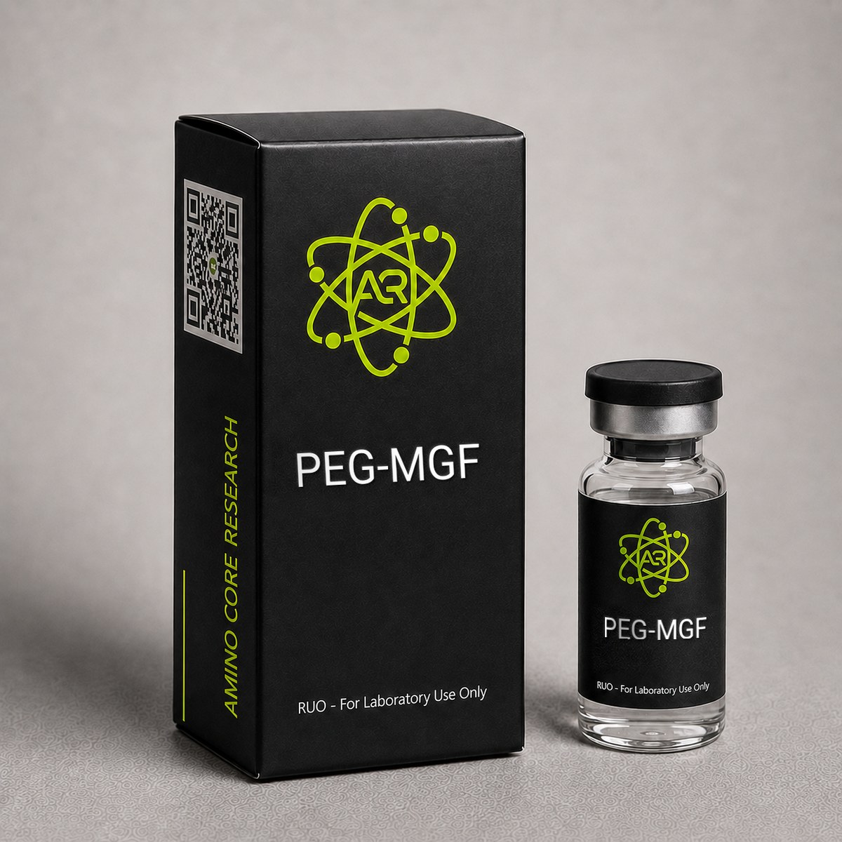
PEG-MGF Peptide
PEGylated form of Mechano Growth Factor with enhanced bioavailability and extended half-life. Studied for skeletal muscle hypertrophy, satellite cell activation, and recovery research.
Research Reference
This page is a research monograph. PEG-MGF is documented here for reference — structure, mechanism and published literature — and is not offered for sale.
Browse compounds available for research purchase · Read the research library
Quick Facts
| SKU | PEGMGF-001 |
|---|---|
| CAS Number | 108174-48-7 |
| Molecular Formula | C121H200N42O39 |
| Molecular Weight | 2867.13 g/mol |
| Sequence | Tyr-Gln-Pro-Pro-Ser-Thr-Asn-Lys-Asn-Thr-Lys-Ser-Gln-Arg-Arg-Lys-Gly-Ser-Thr-Phe-Glu-Glu-Arg-Lys (PEGylated at N-terminus) |
| Purity | ≥99% |
| Physical Form | Lyophilized Powder |
| Storage | Store at -20°C |
What is PEG-MGF?
PEG-MGF (PEGylated Mechano Growth Factor) is a splice variant of IGF-1 with polyethylene glycol modification. The PEG moiety extends the biological half-life from minutes to several hours by reducing renal clearance and proteolytic degradation. MGF (IGF-1Ec) is expressed in response to mechanical stress in muscle tissue and activates satellite cells for muscle repair and hypertrophy.
Mechanism of Action
MGF activates muscle satellite cells (muscle stem cells) via the IGF-1R pathway, promoting their proliferation and fusion into existing myofibers. Unlike systemic IGF-1, MGF acts locally at the site of mechanical damage. The PEG modification allows systemic research administration with sustained satellite cell activation across multiple muscle groups.
Research & Clinical Studies
Local MGF Administration and Satellite Cell Activation
One of the most influential investigations into mechano growth factor (MGF) biology was conducted by Goldspink and colleagues, who demonstrated that MGF — a splice variant of insulin-like growth factor 1 (IGF-1) produced in response to mechanical loading — plays a critical role in initiating muscle repair and hypertrophy. In their landmark study, MGF cDNA was delivered to skeletal muscle of rodents, and the muscles examined for fiber size, satellite cell activation, and myogenic regulatory factor expression.
Study Design
- Model: Adult rodent skeletal muscle (tibialis anterior and extensor digitorum longus)
- Intervention: Intramuscular MGF cDNA delivery or recombinant MGF peptide injection
- Duration: 2-3 weeks post-injection assessment
- Endpoints: Mean fiber cross-sectional area, satellite cell counts, MyoD and Myf5 expression
Key Findings
- +25% increase in mean muscle fiber cross-sectional area within 2 weeks of MGF delivery
- Significant upregulation of MyoD and Myf5, the principal myogenic regulatory factors that drive satellite cell commitment to myogenesis
- Marked satellite cell proliferation, providing additional myonuclei to support fiber hypertrophy
- Effects were localized to injected muscle, supporting MGF's role as an autocrine/paracrine factor distinct from systemic IGF-1
Relevance to PEG-MGF Research
While native MGF has a biological half-life of only a few minutes in circulation, PEGylation extends systemic stability to several days, enabling investigators to model sustained satellite cell signaling without continuous intramuscular injection. PEG-MGF studies have used the findings of Goldspink's group as the mechanistic foundation: if local MGF triggers satellite cell activation, then a long-acting analog should permit systemic study of myogenic regulatory factor induction in models of disuse atrophy, age-related sarcopenia, and post-injury regeneration.
This framework has informed subsequent work in cardiac MGF biology, dystrophic muscle models, and tendon repair research, where PEG-MGF is increasingly used as a tool compound to probe the IGF-1Ec / MGF receptor pathway distinct from canonical IGF-1R signaling.
[1] Goldspink G. Mechanical signals, IGF-I gene splicing, and muscle adaptation. Physiology (Bethesda). 2005;20:232-238. PubMed ↗
[2] Hill M, Goldspink G. Expression and splicing of the insulin-like growth factor gene in rodent muscle is associated with muscle satellite (stem) cell activation following local tissue damage. J Physiol. 2003;549(Pt 2):409-418. PubMed ↗
PEGylation and Pharmacokinetic Extension of MGF
Native MGF (the IGF-1Ec splice variant) is rapidly degraded by serum proteases, with a plasma half-life estimated at less than 5-7 minutes in rodent models. This short window limits its utility for systemic research applications. Polyethylene glycol (PEG) conjugation has been investigated as a strategy to extend circulating half-life of peptide therapeutics, and the same principle has been applied to MGF.
Rationale for PEGylation
PEGylation involves the covalent attachment of one or more PEG chains (typically 5-40 kDa) to a peptide, increasing its hydrodynamic radius, shielding it from proteolytic enzymes, and reducing renal clearance. For MGF, N-terminal PEGylation preserves the C-terminal E-domain sequence that is essential for satellite cell-activating activity, while extending plasma stability.
Reported Pharmacokinetic Profile
- PEG-MGF plasma half-life: ~48-72 hours in preclinical models, compared to ~5 minutes for unmodified MGF
- Sustained serum bioavailability permitting weekly or twice-weekly dosing schedules in research protocols
- Retention of satellite cell activating activity in ex vivo myoblast proliferation assays
- Reduced cross-reactivity with the IGF-1 receptor (IGF-1R) compared to mature IGF-1, suggesting a distinct receptor or splice-variant-specific binding site
Comparative Context
The PEGylation strategy used for MGF parallels approaches successfully applied to interferon-alpha (Pegasys), filgrastim (Neulasta), and growth hormone analogs. In each case, PEG conjugation has converted short-acting peptides into clinically practical long-acting agents. While PEG-MGF remains an investigational research compound and is not approved for human therapeutic use, its pharmacokinetic profile makes it a valuable tool for laboratory investigations into mechano-sensitive growth factor signaling, satellite cell dynamics, and the time-course of myogenic regulatory factor expression in skeletal muscle research models.
Researchers studying PEG-MGF should note that the extended half-life also changes the kinetics of downstream signaling: where native MGF produces a brief, intense pulse of satellite cell activation, PEG-MGF generates a prolonged, lower-amplitude signal that may produce qualitatively different transcriptional profiles. This distinction is an active area of investigation.
[1] Veronese FM, Pasut G. PEGylation, successful approach to drug delivery. Drug Discov Today. 2005;10(21):1451-1458. PubMed ↗
[2] Yang SY, Goldspink G. Different roles of the IGF-I Ec peptide (MGF) and mature IGF-I in myoblast proliferation and differentiation. FEBS Lett. 2002;522(1-3):156-160. PubMed ↗
PEG-MGF and Skeletal Muscle Hypertrophy in Rodent Models
Research investigating PEGylated Mechano Growth Factor (PEG-MGF) in rodent models has examined its capacity to influence skeletal muscle mass, fiber cross-sectional area, and satellite cell dynamics following systemic administration. Because native MGF (the IGF-1Ec splice variant) carries a short circulating half-life of only several minutes, the polyethylene glycol conjugation strategy was developed to enable studies that depend on sustained peripheral exposure. The following observations come from preclinical investigations in mice and rats and should be interpreted strictly within a research context.
Study Design Overview
Goldspink and colleagues, who originally characterized the MGF splice variant, demonstrated in murine models that local injection of MGF cDNA into tibialis anterior muscle produced approximately a 25% increase in muscle mass within three weeks, exceeding the hypertrophy observed with IGF-IEa expression over comparable timeframes [1]. Subsequent PEG-MGF research extended these findings using systemic intramuscular dosing protocols typically in the range of 100-400 µg per animal, administered every 3-5 days over multi-week protocols.
Key Reported Outcomes
- Increased muscle fiber cross-sectional area in treated limbs versus contralateral controls, with reported differences of 15-20% in some short-duration protocols.
- Elevated satellite cell counts identified by Pax7+ immunohistochemistry, consistent with MGF's proposed role in myogenic precursor activation rather than direct stimulation of protein synthesis through the IGF-1R alone.
- Faster recovery from induced muscle damage (cardiotoxin or eccentric exercise models) when PEG-MGF was administered post-injury, with markers such as embryonic myosin heavy chain expression rising earlier than in vehicle controls.
- Sustained plasma exposure across 48-72 hours following a single subcutaneous PEG-MGF dose, compared with rapid clearance of unmodified MGF within minutes.
Mechanistic Interpretation
The hypertrophic response observed in these models is generally attributed to a two-stage mechanism: (1) recruitment and proliferation of quiescent satellite cells via the E-domain peptide signaling through a non-IGF-1R pathway, and (2) downstream IGF-1R/PI3K/Akt/mTOR signaling once the mature IGF-1 domain becomes available. PEGylation appears to preserve the bioactivity of the E-domain while protecting the molecule from rapid proteolytic clearance.
Limitations
Most PEG-MGF data remain preclinical. There are no published Phase 2 or Phase 3 human trials. Interpretation across studies is complicated by variability in PEGylation chemistry (linear vs branched, 20 kDa vs 40 kDa PEG), injection site (intramuscular vs subcutaneous), and species. Researchers should treat reported magnitudes as protocol-specific and not generalizable.
References:
[1] Goldspink G. Mechanical signals, IGF-I gene splicing, and muscle adaptation. Physiology (Bethesda). 2005;20:232-238. PMID: 16024511
[1] Goldspink G. Mechanical signals, IGF-I gene splicing, and muscle adaptation. Physiology (Bethesda). 2005;20:232-238. PubMed ↗
Chemical & Physical Properties
The following table summarizes the verified chemical and physical properties of PEG-MGF (PEGylated Mechano Growth Factor) as used in laboratory research applications:
| Full Name | PEGylated Mechano Growth Factor (PEG-MGF) |
|---|---|
| Synonyms | PEG-MGF, Pegylated IGF-1Ec, Pegylated Mechano Growth Factor |
| Parent Peptide | IGF-1Ec (Mechano Growth Factor, MGF) |
| Molecular Formula (peptide core) | C₁₂₁H₂₀₀N₄₂O₃₉ |
| Molecular Weight (peptide core) | 2,867.13 g/mol |
| Total MW (with PEG) | Variable depending on PEG chain (typically 5-10 kDa added) |
| CAS Number (MGF core) | 108174-48-7 |
| Amino Acid Sequence | Tyr-Gln-Pro-Pro-Ser-Thr-Asn-Lys-Asn-Thr-Lys-Ser-Gln-Arg-Arg-Lys-Gly-Ser-Thr-Phe-Glu-Glu-Arg-Lys |
| Amino Acid Count | 24 residues (C-terminal E-domain of IGF-1Ec) |
| Key Modification | N-terminal polyethylene glycol conjugation |
| Origin / Developer | MGF originally characterized by Goldspink lab (UK); PEGylated variants developed for extended half-life research |
| Physical Form | Lyophilized white powder |
| Solubility | Soluble in bacteriostatic water, sterile water, and 0.9% sodium chloride solution |
| Purity | ≥99% (HPLC) |
| Plasma Half-Life | ~48-72 hours (vs. ~5 min for native MGF) |
| Storage (Lyophilized) | -20°C long-term; 2-8°C short-term |
| Storage (Reconstituted) | 2-8°C for up to 14 days |
PEG-MGF is a research-only compound. The peptide is distinguished from mature IGF-1 by its unique C-terminal E-domain (the "Ec" exon-derived sequence), which is responsible for its satellite cell-activating properties and is not present in systemic IGF-1. The PEGylation modification preserves this active domain while substantially improving pharmacokinetic stability.
Handling & Reconstitution Guidelines
PEG-MGF is supplied as a sterile lyophilized powder. Because the polyethylene glycol moiety is attached to a peptide backbone containing labile peptide bonds and oxidation-sensitive residues, careful reconstitution technique is essential to preserve bioactivity and ensure reproducible results across an experimental protocol.
Recommended Reconstitution Protocol
- Equilibrate the vial to room temperature (20-25°C) for 15-20 minutes before opening. Reconstituting cold lyophilized peptide can cause local condensation and uneven dissolution.
- Select an appropriate diluent. Bacteriostatic water for injection (0.9% benzyl alcohol) is the standard choice for research protocols requiring multi-dose stability. Sterile water for injection or 0.9% sodium chloride may be used for single-use studies.
- Calculate target concentration. A typical reconstitution is 2 mg PEG-MGF + 2 mL diluent = 1 mg/mL. For finer dosing, 2 mg + 1 mL = 2 mg/mL is also common.
- Introduce diluent slowly down the inner wall of the vial — do not inject directly onto the lyophilized cake. This minimizes mechanical disruption of the PEG-peptide conjugate.
- Swirl gently until fully dissolved (typically 1-3 minutes). The solution should appear clear and colorless.
- Do NOT shake or vortex. Mechanical agitation can shear the PEG linkage, introduce micro-aggregates, and denature the peptide domain.
Compound-Specific Handling Notes
- PEG conjugate stability: The PEG-peptide linkage is most stable at near-neutral pH (6.5-7.5). Avoid acidic or strongly alkaline diluents.
- Oxidation sensitivity: The mature IGF-1 domain contains methionine residues susceptible to oxidation. Minimize exposure to air, light, and elevated temperatures.
- Surface adsorption: At very low concentrations (<0.1 mg/mL), PEG-MGF can adsorb to glass and plastic. Use low-binding tubes for dilution series.
- Filtration: If sterile filtration is required post-reconstitution, use a low-protein-binding 0.22 µm PVDF filter and account for minor recovery losses.
Once reconstituted, label the vial with concentration and date. Aliquoting into single-use volumes minimizes freeze-thaw cycles and protects long-term stability for extended studies.
Storage & Stability Information
Proper storage of PEG-MGF is essential to maintain the integrity of both the polyethylene glycol conjugate and the underlying mechano growth factor peptide. The compound is stable as a lyophilized powder under appropriate cold-chain conditions, but reconstituted solutions are significantly more sensitive to thermal, oxidative, and mechanical stress.
Lyophilized Powder Storage
- Long-term (recommended): Store at -20°C in the original sealed vial, protected from light. Under these conditions, lyophilized PEG-MGF is generally stable for 24 months or longer from the date of manufacture.
- Short-term: Storage at 2-8°C (standard laboratory refrigeration) is acceptable for several weeks if -20°C is not available.
- Transit: Brief exposure to ambient room temperature (up to 7 days) during shipping does not appreciably degrade the lyophilized product, owing to the stabilizing effect of the freeze-dried matrix.
Reconstituted Solution Storage
- Refrigerated (2-8°C): Reconstituted PEG-MGF is typically stable for up to 21-28 days when prepared in bacteriostatic water and kept refrigerated, protected from light.
- Frozen aliquots (-20°C or -80°C): For protocols extending beyond one month, prepare single-use aliquots in low-binding microtubes and freeze. Avoid repeated freeze-thaw cycles, which can fragment the PEG linkage and accelerate aggregation.
- Room temperature: Not recommended for reconstituted material. Limit benchtop exposure to the minimum required for dosing.
Compound-Specific Stability Notes
- PEG linker hydrolysis proceeds slowly at neutral pH but accelerates at extremes; maintain solutions near pH 7.
- Methionine oxidation within the IGF-1 domain is the most common degradation pathway — minimize headspace air and use amber vials when possible.
- Aggregation can occur with repeated thermal cycling; visible cloudiness or particulates indicate the solution should be discarded.
- Light sensitivity: Although PEG-MGF is not strongly chromophoric, UV exposure can promote oxidative degradation; store wrapped in foil or in opaque containers.
Researchers should document storage conditions and reconstitution date in laboratory records to support data reproducibility.
Frequently Asked Questions
PEG-MGF vs MGF vs IGF-1 LR3?
MGF is a local muscle growth factor active for minutes. PEG-MGF extends this to hours via PEGylation. IGF-1 LR3 has systemic effects on overall growth. PEG-MGF specifically targets satellite cell activation for muscle repair.
What is PEG-MGF and how is it used in research?
PEG-MGF (Pegylated Mechano Growth Factor) is a synthetic, PEGylated analog of the IGF-1Ec splice variant — a peptide naturally produced in skeletal muscle in response to mechanical loading and injury. The 24-amino-acid C-terminal E-domain sequence is conjugated to polyethylene glycol (PEG) to extend its plasma half-life from approximately 5 minutes (native MGF) to 48-72 hours. In research settings, PEG-MGF is used as a tool compound to investigate satellite cell activation, myogenic regulatory factor (MyoD, Myf5) expression, and skeletal muscle regeneration kinetics in preclinical models of atrophy, injury, and aging.
What is the molecular weight and CAS number of PEG-MGF?
The peptide core of PEG-MGF has a molecular formula of C₁₂₁H₂₀₀N₄₂O₃₉ and a molecular weight of 2,867.13 g/mol. The CAS number for the MGF (IGF-1Ec) parent peptide is 108174-48-7. The total molecular weight of the PEGylated product depends on the size of the PEG chain attached — typically adding 5,000 to 10,000 Da to the peptide core. Purity is standardized at ≥99% by HPLC analysis, and the compound is supplied as a lyophilized white powder for reconstitution.
How should PEG-MGF be stored and reconstituted?
Lyophilized PEG-MGF should be stored at -20°C for long-term stability and 2-8°C for short-term use. For reconstitution, bacteriostatic water or sterile water is added slowly down the side of the vial; the vial is gently swirled (never shaken or vortexed, as this can shear the peptide). A typical working concentration is 2 mg/mL (e.g., 2 mg peptide reconstituted with 1 mL diluent). Once reconstituted, PEG-MGF should be stored at 2-8°C and used within 14 days to preserve bioactivity.
Why does PEG-MGF have a longer half-life than native MGF?
Native MGF is a small peptide that is rapidly cleared from circulation by serum proteases and renal filtration, with a plasma half-life of only about 5 minutes. PEGylation — the covalent attachment of a polyethylene glycol chain to the N-terminus — dramatically increases the molecule's hydrodynamic radius, shielding it from proteolytic enzymes and reducing renal clearance. This extends PEG-MGF's plasma half-life to approximately 48-72 hours in preclinical models, allowing researchers to maintain sustained systemic exposure with infrequent dosing rather than continuous intramuscular delivery.
What sizes of PEG-MGF are available from AminoCore Research?
PEG-MGF is offered by AminoCore Research as a lyophilized powder in standard research vial sizes, typically 2 mg and 5 mg per vial, with bulk quantities available for institutional protocols. Each vial is supplied with a Certificate of Analysis (COA) confirming ≥99% HPLC purity, mass spectrometry confirmation of molecular weight (approximately 2,867 g/mol), and PEGylation verification. All material is intended strictly for in vitro and preclinical laboratory research use and is not for human consumption.
Does PEG-MGF activate satellite cells differently from IGF-1?
Yes. Research suggests that the E-domain of MGF — the C-terminal peptide that distinguishes it from systemic IGF-1Ea — preferentially activates quiescent muscle satellite cells via a signaling pathway that appears to be independent of the classical IGF-1 receptor. In preclinical models, this leads to satellite cell proliferation and entry into the myogenic program, which complements rather than duplicates IGF-1's direct anabolic signaling through PI3K/Akt/mTOR. PEGylation extends the window over which this E-domain signaling can occur by protecting the molecule from rapid proteolytic clearance.
Is PEG-MGF approved for human use?
No. PEG-MGF has not been approved by the FDA, EMA, or any other regulatory authority for human therapeutic use. It is a research compound studied in preclinical models of skeletal muscle adaptation, satellite cell biology, and recovery from induced muscle injury. AminoCore Research supplies PEG-MGF exclusively for in vitro experimentation and laboratory animal research conducted under appropriate institutional oversight. It is not a drug, dietary supplement, or cosmetic product, and it is not intended for diagnosis, treatment, or prevention of any condition in humans.
How does PEGylation affect the bioactivity of MGF?
PEGylation — the covalent attachment of polyethylene glycol chains to the peptide — increases the hydrodynamic radius of MGF, reducing renal filtration and shielding the molecule from circulating proteases. Reported pharmacokinetic studies indicate that PEG-MGF achieves plasma exposure across 48-72 hours following a single dose, compared with minutes for unmodified MGF. The conjugation site and PEG molecular weight (commonly 20-40 kDa) are designed to preserve E-domain accessibility, so satellite cell activation remains the principal mechanism of interest, while extended half-life enables practical dosing intervals in research protocols.
For laboratory and research use only. This product is not for human or veterinary use. It is not a drug, supplement or food.
This product is not FDA approved for any indication, and is not intended to diagnose, treat, cure, or prevent any disease. All product information is derived from published preclinical research and does not constitute medical advice or claims.