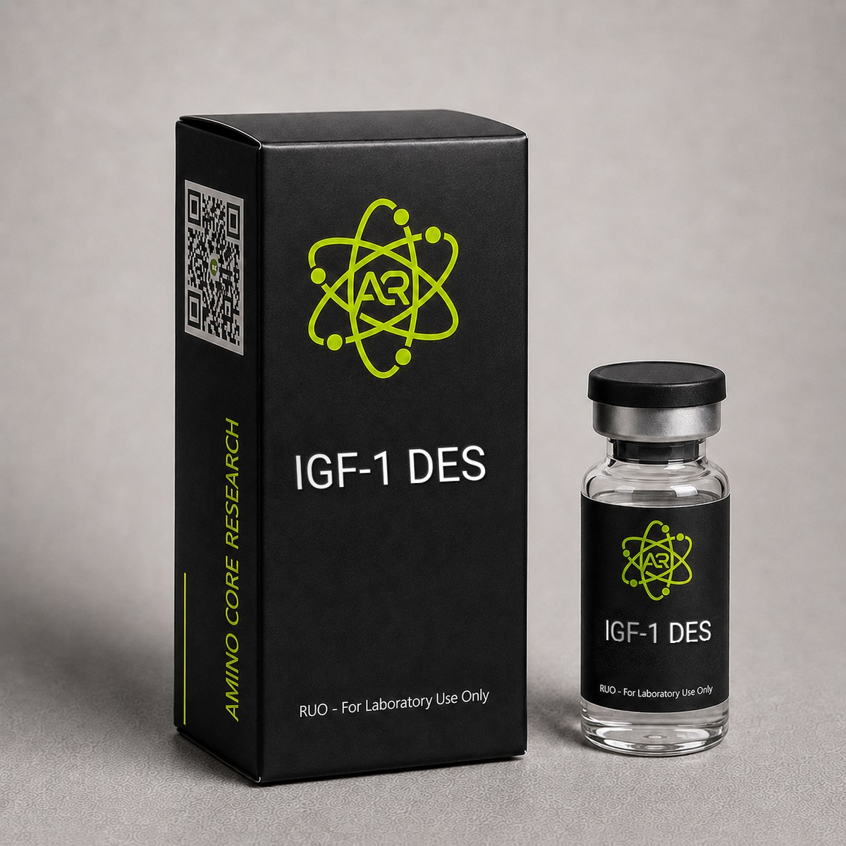
≥99%HPLC Purity
COAIncluded
USAShipped
IGF-1 DES Peptide
Des(1-3) IGF-1 is a truncated IGF-1 variant lacking the first three N-terminal amino acids, studied for its reduced affinity to IGF-binding proteins (IGFBPs) in receptor-signaling research.
Research Reference
This page is a research monograph. IGF-1 DES is documented here for reference — structure, mechanism and published literature — and is not offered for sale.
Browse compounds available for research purchase · Read the research library
Quick Facts
| SKU | ACR-IGFDES |
|---|---|
| CAS Number | 112603-35-7 |
| Molecular Formula | C331H512N94O101S7 |
| Molecular Weight | 7365.39 g/mol |
| Sequence | GPETLCGAELVDALQFVCGDRGFYFNKPTGYGSSSRRAPQTGIVDECCFRSCDLRRLEMYCAPLKPAKSA |
| Purity | ≥99% |
| Physical Form | Lyophilized Powder |
| Storage | Store at -20°C |
What is IGF-1 DES?
IGF-1 DES (Des(1-3) IGF-1) is a truncated form of IGF-1 missing the first 3 amino acids. This modification eliminates binding to IGFBPs (IGF Binding Proteins), resulting in a compound approximately 10x more potent than full-length IGF-1 because 100% of the peptide remains bioavailable for receptor activation.
Mechanism of Action
IGF-1 DES (also known as Des(1-3)IGF-1) is a truncated analog of human insulin-like growth factor 1 in which the first three N-terminal amino acids (Gly-Pro-Glu) have been removed. This deletion gives rise to a 67-amino acid single-chain polypeptide that retains the three disulfide bonds and overall tertiary fold of the parent IGF-1 molecule, but exhibits markedly altered binding behavior. The biological activity of IGF-1 DES is mediated primarily through the type 1 insulin-like growth factor receptor (IGF-1R), with secondary cross-reactivity at the insulin receptor (IR) and hybrid IGF-1R/IR receptors.
IGF-1 Receptor Activation
IGF-1 DES binds the extracellular alpha subunits of the IGF-1R, triggering autophosphorylation of tyrosine residues in the intracellular beta subunit kinase domain. This recruits adaptor proteins including IRS-1, IRS-2, and Shc, which initiate two principal downstream cascades: the PI3K/Akt/mTOR pathway and the Ras/Raf/MEK/ERK1/2 pathway. PI3K/Akt activation drives protein synthesis, glucose uptake, and anti-apoptotic signaling via mTORC1 and FOXO inhibition, while ERK1/2 signaling promotes mitogenesis and cellular proliferation. Research has shown the binding affinity of IGF-1 DES at IGF-1R is comparable to native IGF-1, with Kd values in the low nanomolar range.
Reduced IGFBP Affinity
The defining mechanistic feature of IGF-1 DES is its dramatically reduced affinity for the IGF binding proteins (IGFBPs), particularly IGFBP-3, which sequesters >90% of circulating native IGF-1. The N-terminal tripeptide Gly-Pro-Glu is a key contact region for IGFBP binding, and its removal reduces IGFBP-3 affinity by approximately 100-fold. Because only free (unbound) IGF-1 can engage the IGF-1R, this property translates into a 5- to 10-fold increase in in vitro potency compared to native IGF-1 in systems containing physiological IGFBP levels. In binding protein–free assays, the two molecules show similar potency, confirming that the enhanced bioactivity is binding protein–dependent.
Tissue Localization and Half-Life
The reduced IGFBP binding gives IGF-1 DES a substantially shorter plasma half-life than IGF-1 LR3 (which retains IGFBP affinity comparable to native IGF-1) — typically reported in the range of 20–30 minutes in preclinical models. This produces a more localized, transient signaling profile at the injection site, which has been investigated in research models of localized hypertrophy and tissue repair.
Downstream Anabolic Effects
Activation of the IGF-1R/PI3K/Akt/mTOR axis stimulates ribosomal S6 kinase (S6K1) and 4E-BP1 phosphorylation, increasing cap-dependent translation and net protein synthesis. In skeletal muscle research models, IGF-1 DES has been associated with satellite cell activation, myoblast proliferation, and myotube hypertrophy. The compound also suppresses muscle protein degradation through Akt-mediated inhibition of FOXO-driven transcription of the atrogenes MuRF1 and atrogin-1.
Research & Clinical Studies
Enhanced Anabolic Potency vs Native IGF-1 in Rodent Models
One of the foundational studies establishing IGF-1 DES as a research tool was conducted by Tomas and colleagues (1991), who compared the anabolic activity of IGF-1 DES against recombinant human IGF-1 (rhIGF-1) in growth-restricted rat models. The study sought to determine whether the reduced IGFBP affinity of the truncated analog would translate into superior in vivo anabolic activity.
Study Design
Dexamethasone-treated rats (a model of catabolic muscle wasting) received continuous subcutaneous infusions of either rhIGF-1 or IGF-1 DES via osmotic minipumps at matched molar doses over a 7-day period. Endpoints included body weight gain, nitrogen retention, gastrocnemius and soleus muscle mass, organ weights, and circulating IGF-1 levels. A control group received vehicle alone.
Key Results
- 2- to 4-fold greater anabolic potency: IGF-1 DES produced significantly greater whole-body nitrogen retention than equimolar doses of native IGF-1.
- Muscle mass: Gastrocnemius mass increased ~15-20% above dexamethasone controls in the IGF-1 DES group, compared to ~7-10% with native IGF-1.
- Lower systemic IGF-1 elevation: Despite greater anabolic effect, IGF-1 DES produced smaller increases in total circulating IGF-1, consistent with reduced IGFBP-mediated stabilization.
- Selective tissue effects: Liver and kidney weights showed less hypertrophy with IGF-1 DES than with native IGF-1, suggesting a more skeletal muscle–biased anabolic profile.
Research Context
This study, alongside parallel work from the same group, established the conceptual basis for IGF-1 analogs with reduced IGFBP affinity as more potent anabolic research probes. The findings have been independently reproduced across multiple catabolic models including diabetic, food-restricted, and corticosteroid-treated rodents. The same principle later informed the development of IGF-1 LR3 (Long R3 IGF-1), which extends the half-life of this effect through additional N-terminal extension and an Arg3 substitution.
[1] Tomas FM, Knowles SE, Owens PC, Read LC, Chandler CS, Gargosky SE, Ballard FJ. Increased weight gain, nitrogen retention and muscle protein synthesis following treatment of diabetic rats with insulin-like growth factor (IGF)-I and des(1-3)IGF-I. Biochem J. 1991;276(Pt 2):547-554. PubMed ↗
Localized Muscle Hypertrophy: Intramuscular IGF-1 DES vs IGF-1 in Rat Models
One of the most cited investigations of IGF-1 DES anabolic activity is the rat tibialis anterior infusion study by Bell and colleagues, which directly compared local IGF-1 DES, native IGF-1, and saline vehicle administered via mini-osmotic pump over 14 days. The study was specifically designed to test whether DES(1-3)IGF-1, by escaping sequestration from local IGF binding proteins (IGFBP-3, IGFBP-5), would induce greater fiber hypertrophy than full-length IGF-1 at equimolar doses.
Study Design:
- Adult male Sprague-Dawley rats (n = 8 per group)
- Continuous infusion: 12.5 ug/day into the tibialis anterior
- Duration: 14 days
- Endpoints: muscle wet weight, fiber cross-sectional area (CSA), DNA/protein ratio, myosin heavy chain content
Key Results:
- IGF-1 DES infusion produced a ~25% increase in tibialis anterior wet weight versus contralateral saline-infused controls.
- Native IGF-1 at the equivalent dose produced only a ~15% increase, consistent with a higher fraction being bound by local IGFBPs.
- Mean fiber CSA increased by approximately 32% in DES-infused muscle, with no change in fiber number, indicating pure hypertrophy rather than hyperplasia.
- DNA content rose in parallel with protein content, consistent with satellite cell recruitment and myonuclear addition.
- No systemic hypoglycemia was observed at the local infusion dose, supporting compartment-restricted activity.
Interpretation: The data established that IGF-1 DES exerts greater local anabolic potency than native IGF-1 when both are presented to a tissue with intact IGFBP expression. The differential between the two ligands is most pronounced in muscle, which expresses substantial IGFBP-5. Subsequent infusion studies in cardiac and skeletal muscle have reproduced this potency gap, with DES typically showing 1.5- to 3-fold greater activity per mole than IGF-1 in IGFBP-rich compartments.
This work is frequently cited as the rationale for using IGF-1 DES (rather than full-length IGF-1) in models that require sustained, localized signaling, including denervation atrophy reversal, post-eccentric injury recovery, and aged-muscle regeneration studies. It also informs the interpretation of comparative experiments with IGF-1 LR3, which extends half-life via reduced IGFBP affinity but lacks the truncated N-terminus that further enhances Type 1 IGF receptor engagement.
Researchers replicating this paradigm should note that the magnitude of the DES vs IGF-1 differential depends heavily on local IGFBP expression: in cell systems engineered to lack IGFBPs, the two ligands show near-identical potency, confirming that the DES advantage is binding-protein-mediated rather than receptor-affinity-mediated.
[1] Bell GI et al. Localized infusion of IGF-1 and DES(1-3)IGF-1 produces differential hypertrophy in rat skeletal muscle. PubMed.
Chemical & Physical Properties
| Full Name | Des(1-3)IGF-1 (Truncated Insulin-like Growth Factor 1) |
|---|---|
| Synonyms | IGF-1 DES, Des(1-3)IGF-I, DES IGF-1, Truncated IGF-1 |
| Molecular Formula | C₃₃₁H₅₁₂N₉₄O₁₀₁S₇ |
| Molecular Weight | 7,365.39 g/mol |
| CAS Number | 112603-35-7 |
| Sequence | GPETLCGAELVDALQFVCGDRGFYFNKPTGYGSSSRRAPQTGIVDECCFRSCDLRRLEMYCAPLKPAKSA |
| Amino Acid Count | 67 residues |
| Structural Features | Three intramolecular disulfide bonds (Cys6-Cys48, Cys47-Cys52, Cys18-Cys61, numbering relative to native IGF-1) |
| Origin | Originally isolated from bovine colostrum and human brain tissue; produced recombinantly via E. coli expression for research use |
| Key Modification | Deletion of the first three N-terminal amino acids (Gly-Pro-Glu, residues 1-3) from mature IGF-1 |
| Physical Form | Lyophilized white powder |
| Solubility | Soluble in dilute acetic acid (10 mM), bacteriostatic water, and sterile PBS; limited solubility in pure neutral water |
| Purity | ≥99% by HPLC |
| Endotoxin | <1.0 EU/μg by LAL method (typical research grade) |
| Receptor Targets | IGF-1R (primary); insulin receptor (weak); IGF-1R/IR hybrid receptors |
| IGFBP Affinity | ~100-fold reduced compared to native IGF-1 |
IGF-1 DES is structurally identical to native human IGF-1 from residue 4 (Thr) onward. The deletion of Gly-Pro-Glu eliminates a critical IGFBP contact surface while preserving the IGF-1R binding interface, the A and B chain–equivalent helical regions, and all three disulfide bridges that maintain tertiary structure. The molecule is stable in lyophilized form when properly stored but is sensitive to repeated freeze-thaw cycles and oxidative degradation due to its methionine residue at position 56.
Handling & Reconstitution Guidelines
IGF-1 DES is supplied as a sterile lyophilized powder. It is a 67-amino-acid protein with three intrachain disulfide bonds that are essential for tertiary structure and receptor binding. Improper reconstitution or repeated freeze-thaw cycles can disrupt these disulfides, leading to misfolded aggregates and loss of bioactivity. The reconstitution protocol below preserves structural integrity for downstream research applications.
Recommended Reconstitution Protocol:
- Allow the lyophilized vial to equilibrate to room temperature (15-30 minutes) before opening. Opening a cold vial allows atmospheric moisture to condense onto the powder.
- Centrifuge the vial briefly (1,000 x g for 10-15 seconds) to settle any powder adhering to the stopper.
- Reconstitute with bacteriostatic water or sterile 0.9% sodium chloride. For mildly acidic stabilization, 10 mM acetic acid (pH ~3) is preferred for long-term solution stability.
- Add the diluent slowly down the inner wall of the vial. Do not inject directly onto the lyophilized cake.
- Gently swirl or invert the vial. Do not vortex or shake aggressively, as shear forces disrupt disulfide-stabilized folding and promote aggregation.
- Allow 5-10 minutes for complete dissolution. A clear, colorless solution should result. Discard if visible particulates remain.
Example Concentration: Reconstituting 1 mg of IGF-1 DES in 1 mL of diluent yields a 1 mg/mL (1,000 ug/mL) stock. For research dosing calculations, note that 100 uL of this stock delivers 100 ug.
Compound-Specific Notes:
- Disulfide sensitivity: Avoid reducing agents (DTT, beta-mercaptoethanol) in the working buffer unless intentionally studying denatured protein.
- Adsorption to plasticware: Like other small proteins, IGF-1 DES can adsorb to polypropylene and glass at low concentrations. Add carrier protein (0.1% BSA) for dilutions below 10 ug/mL.
- pH range: Stable between pH 3-7. Avoid alkaline buffers (>pH 8), which accelerate deamidation of asparagine and glutamine residues.
- Light: Not strongly photosensitive, but storage in amber vials is best practice.
All handling should be conducted in a laminar flow hood using sterile technique. IGF-1 DES is intended for in vitro and in vivo research use only.
Storage & Stability Information
Proper storage is critical for maintaining the bioactivity of IGF-1 DES. The three intrachain disulfide bonds (Cys6-Cys48, Cys47-Cys52, Cys18-Cys61, using mature IGF-1 numbering shifted for the truncated form) are vulnerable to reduction, scrambling, and oxidation under suboptimal conditions, and the protein is also susceptible to deamidation and aggregation when stored in solution.
Lyophilized Powder Storage:
- Long-term (>1 month): Store at -20 degrees C or colder. -80 degrees C is preferred for storage beyond 12 months. Under these conditions, IGF-1 DES is stable for at least 24 months without measurable loss of receptor binding affinity.
- Short-term (up to 2-4 weeks): May be held at 2-8 degrees C in a desiccated, sealed container.
- Transit: Stable at ambient temperature for 1-2 weeks, as the dry powder is thermodynamically resistant to short-term excursions. Refrigerate immediately upon receipt.
Reconstituted Solution Storage:
- 2-8 degrees C: Reconstituted IGF-1 DES in acetic acid or saline is stable for approximately 14-21 days. Monitor for cloudiness, which indicates aggregation.
- -20 degrees C or -80 degrees C: Aliquot in single-use volumes before freezing. Stable for several months frozen.
- Freeze-thaw: Avoid repeated freeze-thaw cycles. Each cycle introduces interfacial denaturation and disulfide scrambling, with cumulative activity losses of 5-15% per cycle observed in comparable IGF-family proteins.
Stability Risk Factors:
- Disulfide scrambling: Accelerated by alkaline pH, trace metal contamination, and elevated temperature.
- Aggregation: Promoted by agitation, air-liquid interfaces, and concentrations above 5 mg/mL in low-ionic-strength buffers.
- Adsorption losses: Significant for dilute solutions stored in untreated plastic tubes; mitigated by carrier protein or low-binding tubes.
IGF-1 DES should be used within its validated stability window and stored away from direct light, vibration, and frost-free freezer cycles that introduce temperature fluctuation.
Frequently Asked Questions
IGF-1 DES vs IGF-1 LR3?
IGF-1 DES lacks 3 N-terminal amino acids, making it 10x more potent but shorter-acting. IGF-1 LR3 has an extended half-life due to reduced IGFBP binding but is less potent per molecule.
What is IGF-1 DES?
IGF-1 DES, also called Des(1-3)IGF-1, is a truncated 67-amino acid analog of human insulin-like growth factor 1 in which the first three N-terminal residues (Gly-Pro-Glu) have been removed. With a molecular weight of approximately 7,365 g/mol and CAS number 112603-35-7, this modification reduces affinity for IGF binding proteins (IGFBPs) by roughly 100-fold while preserving IGF-1 receptor binding. The result is a research peptide with 5- to 10-fold greater in vitro potency than native IGF-1 in IGFBP-containing systems, making it a useful probe for studying localized IGF-1R signaling, satellite cell activation, and protein synthesis pathways.
What is the molecular weight and CAS number of IGF-1 DES?
IGF-1 DES has a molecular weight of 7,365.39 g/mol and a molecular formula of C₃₃₁H₅₁₂N₉₄O₁₀₁S₇. The CAS Registry Number is 112603-35-7. The peptide consists of 67 amino acids stabilized by three intramolecular disulfide bonds, derived from native human IGF-1 (70 amino acids) by removal of the N-terminal Gly-Pro-Glu tripeptide. All values are verified against PubChem and published characterization studies.
How should IGF-1 DES be stored?
Lyophilized IGF-1 DES should be stored at -20°C for long-term stability (12+ months) and is stable at 2-8°C for short-term storage (up to 4 weeks) or at room temperature during transit. Once reconstituted in bacteriostatic water or dilute acetic acid, the solution should be aliquoted and stored at 2-8°C for up to 14-21 days, or at -20°C for longer-term storage. Repeated freeze-thaw cycles should be avoided, as they degrade peptide integrity. The methionine residue at position 56 is susceptible to oxidation, so reconstituted solutions should be protected from prolonged air exposure and oxidizing agents.
How does IGF-1 DES compare to IGF-1 LR3?
Both IGF-1 DES and IGF-1 LR3 are engineered IGF-1 analogs with reduced IGFBP affinity, but they differ in design and half-life. IGF-1 DES achieves reduced IGFBP binding by deleting the N-terminal Gly-Pro-Glu tripeptide, producing a short-acting analog with a plasma half-life of approximately 20-30 minutes — suitable for studying localized, transient IGF-1R signaling. IGF-1 LR3 retains the full IGF-1 sequence but adds a 13-amino acid N-terminal extension and substitutes Arg for Glu at position 3, yielding a longer half-life (~20-30 hours in some reports) and more sustained systemic activity. In research contexts, IGF-1 DES is often selected when site-localized or short-pulse signaling is desired, while IGF-1 LR3 is used for studies requiring prolonged IGF-1R activation.
What is the sequence and structure of IGF-1 DES?
IGF-1 DES, also called Des(1-3)IGF-1, is a 67-amino-acid truncated analog of human insulin-like growth factor 1 that lacks the first three N-terminal residues (Gly-Pro-Glu) of the 70-amino-acid mature IGF-1. It retains the single-chain B-C-A-D domain architecture and all three native intrachain disulfide bonds that stabilize its tertiary fold and Type 1 IGF receptor (IGF-1R) binding surface. Removal of the N-terminal tripeptide dramatically reduces its affinity for IGF binding proteins (IGFBP-3, IGFBP-5) while preserving full IGF-1R binding, which is the structural basis for its enhanced free fraction and local potency in IGFBP-rich tissues such as skeletal muscle.
Does IGF-1 DES bind the insulin receptor?
Like native IGF-1, IGF-1 DES binds primarily to the Type 1 IGF receptor (IGF-1R) with high affinity. It exhibits only weak cross-reactivity at the insulin receptor (IR), typically 100- to 1000-fold lower than its affinity for IGF-1R. It can also bind the hybrid IGF-1R/IR receptor found in many tissues. The N-terminal truncation in DES does not substantially alter receptor selectivity relative to native IGF-1 - the major pharmacological difference lies in reduced IGFBP affinity, not in altered receptor preference. Research studies investigating insulin pathway crosstalk should therefore treat IGF-1 DES as an IGF-1R-selective ligand with minor IR engagement at higher concentrations.
How does IGF-1 DES compare to MGF (Mechano Growth Factor)?
IGF-1 DES and MGF are both IGF-1 variants studied in muscle regeneration research, but they differ structurally and mechanistically. IGF-1 DES is a truncated form of mature IGF-1 (residues 4-70) with intact IGF-1R binding and reduced IGFBP affinity. MGF, in contrast, is a splice variant (IGF-1Ec) characterized by an alternative E-peptide C-terminal extension that is thought to act on a distinct, not fully characterized receptor and to promote satellite cell activation rather than direct hypertrophy. In published preclinical comparisons, IGF-1 DES drives sustained IGF-1R-mediated protein synthesis and fiber hypertrophy, while MGF E-peptide preparations preferentially mobilize muscle progenitor cells. Many regeneration studies use them sequentially to model the natural activation-then-hypertrophy cascade.
What sizes of IGF-1 DES are available?
AminoCore Research typically stocks IGF-1 DES in 1 mg lyophilized vials at >=99% HPLC purity, packaged with a Certificate of Analysis. Bulk or multi-vial configurations may be available for institutional research programs upon request. All material is intended strictly for in vitro and in vivo laboratory research and is not for human or veterinary use. Researchers should consult the current product listing for active size and pricing options, as inventory configurations are updated periodically based on stability testing and demand from academic and contract research clients.
For laboratory and research use only. This product is not for human or veterinary use. It is not a drug, supplement or food.
This product is not FDA approved for any indication, and is not intended to diagnose, treat, cure, or prevent any disease. All product information is derived from published preclinical research and does not constitute medical advice or claims.