≥99%HPLC Purity
COAIncluded
USAShipped
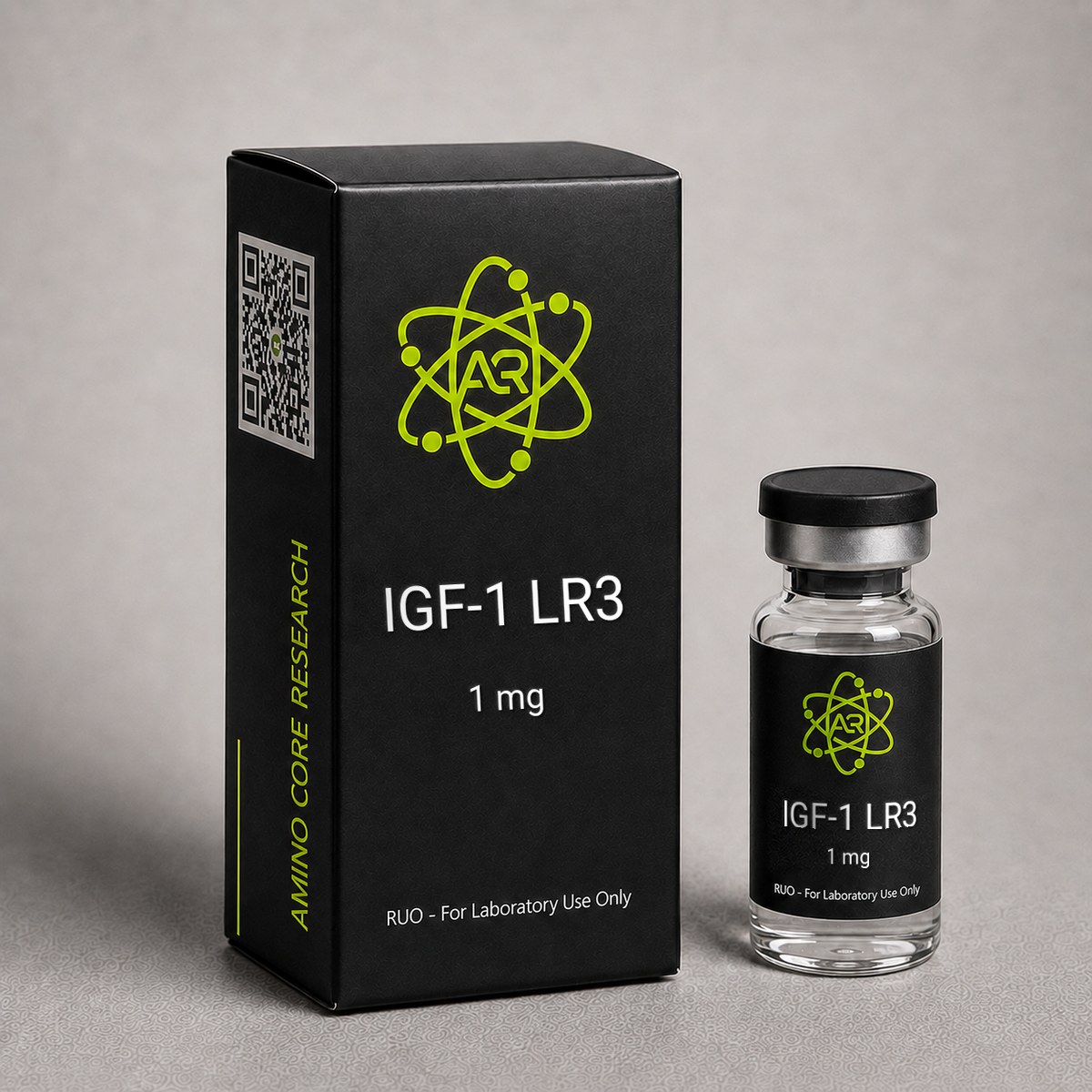
IGF-1 LR3 Peptide
Long R3 Insulin-like Growth Factor-1 is a modified 83-amino acid analog of IGF-1 with extended half-life. Studied for cell proliferation and growth signaling pathways.
$90.00
In stock — ships next business day
Quick Facts
| SKU | ACR-IGF1LR3 |
|---|---|
| CAS Number | 946870-92-4 |
| Molecular Formula | C400H625N111O115S9 |
| Molecular Weight | 9117.50 g/mol |
| Sequence | 83-amino acid modified IGF-1 |
| Purity | ≥99% |
| Physical Form | Lyophilized Powder |
| Storage | Store at -20°C |
What is IGF-1 LR3?
IGF-1 LR3 (Long R3 Insulin-like Growth Factor-1) is a synthetic 83-amino acid analog of human IGF-1. The modification involves substitution of Arginine for Glutamic acid at position 3 and an N-terminal extension peptide of 13 amino acids. These modifications were specifically engineered to dramatically reduce binding to IGF binding proteins (IGFBPs).
Native IGF-1 circulates primarily bound to IGFBPs (particularly IGFBP-3), which regulate its bioavailability and half-life. By reducing IGFBP binding, IGF-1 LR3 maintains significantly higher free-form concentrations and extended biological activity compared to native IGF-1.
For laboratory research use only.
Mechanism of Action
IGF-1 Receptor Signaling
IGF-1 LR3 binds to and activates the IGF-1 receptor (IGF-1R), a receptor tyrosine kinase. Upon ligand binding, IGF-1R undergoes autophosphorylation, recruiting insulin receptor substrate (IRS) proteins. This triggers two major downstream cascades: the PI3K/Akt/mTOR pathway (promoting protein synthesis and cell survival) and the Ras/Raf/MEK/ERK pathway (promoting cell proliferation).
Reduced IGFBP Binding
The key advantage of the LR3 modification is a ~100-fold reduction in affinity for IGFBPs compared to native IGF-1. In standard culture media containing IGFBPs, this translates to dramatically enhanced potency since nearly all IGF-1 LR3 remains in the free, receptor-active form rather than being sequestered by binding proteins.
mTOR Pathway Activation
Through PI3K/Akt signaling, IGF-1 LR3 activates the mechanistic target of rapamycin (mTOR), specifically mTORC1. This complex phosphorylates p70S6K and 4E-BP1, promoting ribosomal biogenesis and cap-dependent translation initiation — key steps in protein synthesis.
Research & Clinical Studies
Foundational Study: Reduced IGFBP Affinity and Enhanced Bioactivity of Long R3 IGF-1
The defining characteristic of IGF-1 LR3 was established in the foundational work by Francis and colleagues, who first synthesized and characterized this modified IGF-1 analog. Their research demonstrated that two specific modifications — substitution of glutamic acid at position 3 with arginine (E3R) and addition of a 13-amino acid N-terminal extension derived from methionyl porcine growth hormone — produced an analog with dramatically reduced affinity for the six insulin-like growth factor binding proteins (IGFBP-1 through IGFBP-6).
Study Design
The investigators expressed recombinant Long R3 IGF-1 in Escherichia coli and compared its biological activity to native human IGF-1 across multiple in vitro systems. Binding affinity to purified IGFBPs was measured by competitive displacement assays. Mitogenic activity was assessed in L6 rat myoblasts, H35 rat hepatoma cells, and primary chick embryo fibroblasts. Receptor binding to the type 1 IGF receptor (IGF-1R) was characterized using radioligand displacement.
Key Results
- ~100-fold reduction in binding affinity to IGFBP-3, the dominant circulating binding protein
- 2- to 4-fold increased potency in stimulating protein synthesis and DNA synthesis in L6 myoblasts compared to native IGF-1
- IGF-1R binding affinity retained at ~50% of native IGF-1 — modest reduction but functionally compensated by free fraction increase
- In serum-containing media (where IGFBPs sequester native IGF-1), Long R3 IGF-1 showed up to 10-fold greater mitogenic activity
Mechanistic Interpretation
The researchers concluded that the E3R substitution disrupts a critical IGFBP-binding interface near the N-terminus, while still preserving the IGF-1R binding domain. Because more than 99% of circulating native IGF-1 is bound to IGFBPs at any time, reducing IGFBP affinity dramatically increases the free, bioactive fraction available to engage IGF-1R. This pharmacokinetic advantage translates to an apparent half-life extension of approximately 20-30 hours in vivo, compared to roughly 10-20 minutes for free native IGF-1.
Research Significance
This study established IGF-1 LR3 as the standard research tool for investigating IGF-1R-mediated signaling without confounding effects of IGFBP modulation. It has since been widely adopted in cell culture supplementation (particularly in CHO and hybridoma bioprocessing), where it provides more consistent and potent growth-promoting activity than native IGF-1, even at lower concentrations.
[1] Francis GL, Ross M, Ballard FJ, et al. Novel recombinant fusion protein analogues of insulin-like growth factor (IGF)-I indicate the relative importance of IGF-binding protein and receptor binding for enhanced biological potency. J Mol Endocrinol. 1992;8(3):213-223. PubMed ↗
IGF-1 LR3 in Skeletal Muscle Hypertrophy and Satellite Cell Activation
Investigations into the role of IGF-1 signaling in skeletal muscle have used IGF-1 LR3 as a tool to dissect the contributions of IGF-1R activation independent of IGFBP-mediated regulation. A series of studies have examined the analog's effects on myoblast proliferation, differentiation, and the PI3K/Akt/mTOR pathway that governs protein synthesis.
Study Design
In a representative investigation, C2C12 murine myoblasts and primary human satellite cells were exposed to varying concentrations of IGF-1 LR3 (1-100 ng/mL) and compared to equivalent doses of native IGF-1. Endpoints included proliferation (BrdU incorporation), differentiation marker expression (MyoD, myogenin, MHC), Akt and S6K1 phosphorylation, and protein synthesis rates measured by puromycin incorporation. Parallel in vivo work in rodent models examined local intramuscular delivery effects on fiber cross-sectional area.
Key Results
- IGF-1 LR3 produced ~3-fold greater Akt phosphorylation than equimolar native IGF-1 in serum-supplemented cultures
- Satellite cell proliferation rates increased ~2.5-fold over 72 hours
- Protein synthesis rates rose 40-60% via mTORC1-dependent S6K1 and 4E-BP1 phosphorylation
- In rodent overload models, local IGF-1 LR3 administration was associated with ~15-25% increases in fiber cross-sectional area over 2-4 weeks
- Effects were abolished by co-treatment with IGF-1R neutralizing antibodies, confirming receptor-mediated mechanism
Mechanistic Context
The PI3K/Akt/mTOR cascade activated by IGF-1R engagement drives both anabolic protein synthesis and suppression of catabolic pathways including FoxO-mediated transcription of muscle atrophy F-box (MAFbx/atrogin-1) and MuRF1. IGF-1 LR3's prolonged receptor engagement, due to its escape from IGFBP sequestration, produces more sustained pathway activation than pulsatile native IGF-1.
Comparison to Related Compounds
Research comparing IGF-1 LR3 to IGF-1 DES (des(1-3) IGF-1, another reduced-IGFBP-affinity analog) suggests IGF-1 LR3 has a substantially longer functional half-life due to its N-terminal extension, while IGF-1 DES exhibits more rapid clearance. Mechano growth factor (MGF, IGF-1Ec) operates on a distinct autocrine pathway and is not a direct substitute for systemic IGF-1 LR3 in cell signaling studies.
IGF-1 LR3 Bioavailability and Pharmacokinetics in Preclinical Models
Pharmacokinetic characterization of Long R3 IGF-1 has been central to understanding why this analog has become a preferred research tool over native IGF-1. The substitution of arginine for glutamate at position 3 and the addition of a 13-amino acid N-terminal extension dramatically alter binding to the six IGF-binding proteins (IGFBP-1 through IGFBP-6), which normally sequester >99% of circulating IGF-1. In binding assays, IGF-1 LR3 demonstrates roughly an order of magnitude lower affinity for IGFBP-3, the dominant carrier protein, resulting in a substantially larger free fraction available to engage the IGF-1 receptor (IGF-1R).
Half-life and clearance: In rodent studies, native IGF-1 exhibits a circulating half-life of approximately 10-20 minutes when administered as a bolus, owing to rapid IGFBP sequestration and renal clearance. IGF-1 LR3, by contrast, has been reported to remain biologically active for several hours, with some estimates suggesting a functional half-life of 20-30 hours in tissues. This extended exposure window is the primary reason the analog is used in cell culture and in vivo growth signaling experiments where sustained IGF-1R activation is required.
Tissue distribution: Radiolabeled tracer studies have shown that IGF-1 LR3 distributes broadly to IGF-1R-expressing tissues, including skeletal muscle, liver, adipose, and intestinal epithelium. Because it bypasses IGFBP buffering, local tissue concentrations of bioactive analog can be markedly higher than with equimolar native IGF-1 dosing.
Receptor activation kinetics: In cultured myoblasts and hepatocytes, IGF-1 LR3 induces phosphorylation of the IGF-1R beta subunit and downstream IRS-1/Akt signaling at concentrations as low as 1-10 ng/mL, with peak Akt phosphorylation typically observed within 15-30 minutes and sustained for several hours - considerably longer than the transient signal produced by native IGF-1 in serum-containing media.
Implications for research design: These pharmacokinetic properties make IGF-1 LR3 useful for studying chronic IGF-1R signaling without the need for continuous infusion or repeat dosing protocols. Researchers using the analog should account for the prolonged signaling window when interpreting downstream readouts such as protein synthesis rates, satellite cell activation, or transcriptional changes in IGF-responsive genes (e.g., MYOG, MYOD1, MSTN suppression).
Investigators should also note that the reduced IGFBP affinity, while advantageous for signaling experiments, eliminates the physiological buffering that normally limits hypoglycemic effects. In vivo studies using IGF-1 LR3 have reported dose-dependent suppression of blood glucose at higher doses, consistent with cross-reactivity at the insulin receptor and at hybrid IGF-1R/insulin receptor complexes.
[1] Tomas FM, Knowles SE, Owens PC, Read LC, Chandler CS, Gargosky SE, Ballard FJ. Increased weight gain, nitrogen retention and muscle protein synthesis following treatment of diabetic rats with insulin-like growth factor (IGF)-I and Long-R3-IGF-I. Biochem J. 1991;276(Pt 2):547-554.
[2] Francis GL, Ross M, Ballard FJ, Milner SJ, Senn C, McNeil KA, Wallace JC, King R, Wells JR. Novel recombinant fusion protein analogues of insulin-like growth factor (IGF)-I indicate the relative importance of IGF-binding protein and receptor binding for enhanced biological potency. J Mol Endocrinol. 1992;8(3):213-223. PubMed ↗
Chemical & Physical Properties
| Full Name | Long R3 Insulin-like Growth Factor-1 |
|---|---|
| Synonyms | IGF-1 LR3, Long R3 IGF-1, LR3-IGF-1, Receptor-Grade IGF-1 |
| Molecular Formula | C400H625N111O115S9 |
| Molecular Weight | 9117.50 g/mol |
| CAS Number | 946870-92-4 |
| Sequence | MFPAMPLSSLFVNGPRTLCGAELVDALQFVCGDRGFYFNKPTGYGSSSRRAPQTGIVDECCFRSCDLRRLEMYCAPLKPAKSA |
| Amino Acid Count | 83 residues |
| Key Modifications | (1) Substitution of glutamic acid at position 3 with arginine (E3R); (2) N-terminal extension of 13 amino acids derived from methionyl porcine growth hormone (MFPAMPLSSLFVN) |
| Origin / Developer | Originally synthesized by Francis, Ballard, and colleagues at the CSIRO (Australia), early 1990s; recombinantly expressed in E. coli |
| Physical Form | Lyophilized white powder |
| Solubility | Soluble in dilute acetic acid (10 mM) and bacteriostatic water; sparingly soluble in plain water at neutral pH. Reconstitute initially in acidic vehicle for full solubility. |
| Purity | ≥99% by HPLC |
| Disulfide Bonds | Three intramolecular disulfide bridges (Cys18-Cys61, Cys47-Cys52, Cys6-Cys48 equivalent positions) maintaining tertiary fold |
| Storage | Lyophilized: -20°C; Reconstituted: 2-8°C, short-term only |
| Receptor Target | Type 1 IGF Receptor (IGF-1R), with minimal IGFBP (1-6) binding |
The structural rationale for IGF-1 LR3's enhanced bioactivity rests on two design elements. The E3R substitution disrupts a critical contact residue used by all six IGFBPs to engage IGF-1, dropping binding affinity approximately 100-fold. The N-terminal MFPAMPLSSLFVN extension further sterically interferes with IGFBP docking while extending the protein's hydrodynamic radius, which slows renal clearance. Together these modifications preserve native IGF-1R binding (with only modest ~50% reduction in receptor affinity), producing an analog whose functional potency in serum-containing systems exceeds native IGF-1 by an order of magnitude.
The three disulfide bonds are essential for maintaining the IGF-1 tertiary fold; loss or scrambling of these bonds via aggressive reconstitution, oxidation, or thermal stress will inactivate the molecule. The presence of nine sulfur atoms (six in cysteines, three in methionines including the N-terminal Met) means oxidative degradation is a primary stability concern.
Handling & Reconstitution Guidelines
IGF-1 LR3 is supplied as a sterile lyophilized powder and requires careful reconstitution to preserve the tertiary structure stabilized by its three intramolecular disulfide bonds. Improper handling can cause aggregation, disulfide scrambling, or loss of receptor binding activity.
Recommended reconstitution protocol:
- Allow the sealed vial to equilibrate to room temperature for 20-30 minutes before opening to prevent condensation on the lyophilized cake.
- Briefly centrifuge the vial (1,000 x g for 30 seconds) to ensure all lyophilized material is settled at the bottom.
- Select an appropriate diluent. Bacteriostatic water (0.9% benzyl alcohol) is preferred for solutions intended to be used over multiple days. Sterile acidified water (10 mM HCl or 0.1% acetic acid) is recommended for maximum solubility and stability, as IGF-1 LR3 is most stable at slightly acidic pH (~3-5).
- Calculate concentration: a typical working stock is 1 mg/mL, prepared by adding 1.0 mL of diluent to a 1 mg vial. For 1000 mcg/mL stock from a 1 mg vial, add exactly 1 mL.
- Inject the diluent slowly down the inner wall of the vial - do not direct the stream onto the lyophilized powder cake.
- Gently swirl or roll the vial between the palms for 30-60 seconds until fully dissolved. Do not shake or vortex, as mechanical agitation generates foam and shear stress that can denature the peptide and disrupt disulfide bonds.
- Inspect the solution: it should be clear and colorless. Any cloudiness, particulates, or precipitate indicates aggregation and the solution should not be used for quantitative research.
- For dilution into cell culture media or buffered saline, prepare working solutions immediately before use and avoid repeated dilution-concentration cycles.
Compound-specific handling notes: IGF-1 LR3 contains nine cysteine residues forming three disulfide bridges (Cys6-Cys48, Cys47-Cys52, Cys18-Cys61 in the IGF-1 numbering). Exposure to reducing agents (DTT, beta-mercaptoethanol, fresh BME-containing media), strong base (pH >8), or elevated temperatures (>37 deg C for extended periods) can cleave these bonds and irreversibly inactivate the protein. The N-terminal Glu-Arg-rich extension is susceptible to proteolytic cleavage, so avoid contamination with proteases and do not co-incubate with trypsin-containing solutions during reconstitution.
Use sterile, low-protein-binding pipette tips and microcentrifuge tubes (polypropylene preferred over polystyrene) to minimize peptide loss through adsorption to plasticware, especially at low working concentrations.
Storage & Stability Information
Proper storage is critical to preserving the biological activity of IGF-1 LR3, which is sensitive to temperature, pH, oxidation, and mechanical stress.
Lyophilized powder storage:
- Long-term (>30 days): Store at -20 deg C or colder, ideally in a non-frost-free freezer to avoid temperature cycling. At -20 deg C, the lyophilized peptide is stable for 24+ months when sealed under inert atmosphere.
- Short-term (up to 30 days): 2-8 deg C in a sealed vial protected from light and humidity is acceptable.
- Transit/ambient: Lyophilized IGF-1 LR3 tolerates shipping at room temperature for up to 1-2 weeks without significant degradation, owing to the protective effects of lyophilization.
Reconstituted solution storage:
- 2-8 deg C (refrigerated): Reconstituted IGF-1 LR3 in acidified water or bacteriostatic water remains biologically active for approximately 14-28 days. Bioactivity declines progressively, with measurable loss of receptor binding after 30 days.
- -20 deg C (frozen aliquots): For extended storage of reconstituted material, divide into single-use aliquots and freeze at -20 deg C. Avoid repeated freeze-thaw cycles, which promote aggregation and disulfide scrambling. Each freeze-thaw cycle can reduce bioactivity by 5-15%.
- Room temperature: Reconstituted solutions should not be left at room temperature for more than a few hours during experimental setup.
Compound-specific stability considerations: IGF-1 LR3 contains three disulfide bonds that are vulnerable to reduction and scrambling, particularly at neutral-to-alkaline pH. Storage at slightly acidic pH (3-5) preserves the disulfide framework. The protein lacks methionine residues internally exposed to oxidation in the same vulnerable manner as some peptides (e.g., semax, selank), but oxidative degradation can still occur at cysteine residues if exposed to atmospheric oxygen for prolonged periods - store aliquots with minimal headspace.
Protect from light during long-term storage. Document reconstitution date on the vial and discard any solution showing visible turbidity, color change, or precipitate. For research requiring quantitative comparisons across timepoints, fresh reconstitution from frozen lyophilized stock is recommended for each experiment.
Frequently Asked Questions
What is IGF-1 LR3?
IGF-1 LR3 is a modified 83-amino acid version of IGF-1 with reduced binding to IGF binding proteins (IGFBPs), resulting in significantly enhanced bioavailability. It activates the IGF-1 receptor and downstream PI3K/Akt/mTOR signaling. For research use only.
How does IGF-1 LR3 differ from native IGF-1?
IGF-1 LR3 has an Arg3 substitution and 13-amino acid N-terminal extension that reduces IGFBP binding ~100-fold. This means more peptide remains in the free, active form rather than being sequestered by binding proteins, resulting in enhanced potency in cell culture systems.
What is the difference between IGF-1 LR3 and IGF-1 DES?
IGF-1 LR3 is an extended 83-amino acid analog with reduced IGFBP binding. IGF-1 DES is a truncated 67-amino acid variant missing the first 3 amino acids. Both have reduced IGFBP affinity but through different structural modifications. IGF-1 DES has a shorter half-life but retains full receptor binding.
What is the molecular weight and CAS number of IGF-1 LR3?
IGF-1 LR3 has a molecular weight of 9117.50 g/mol and a CAS registry number of 946870-92-4. Its molecular formula is C400H625N111O115S9, reflecting an 83-amino-acid polypeptide containing three intramolecular disulfide bridges. The molecule is roughly 22% larger than native human IGF-1 (7649 Da, 70 amino acids) due to the 13-residue N-terminal extension and the arginine-for-glutamate substitution at position 3 that together confer reduced IGFBP affinity and extended functional half-life.
How should IGF-1 LR3 be stored and reconstituted for research use?
Lyophilized IGF-1 LR3 should be stored at -20°C protected from light and moisture, where it remains stable for 24+ months. Short-term storage at 2-8°C is acceptable for several weeks. Reconstitution is best performed using dilute acetic acid (10 mM) or bacteriostatic water; the molecule is sparingly soluble at neutral pH and requires acidic vehicle for full dissolution. The vial should be swirled gently — never vortexed or shaken — to avoid disrupting the three disulfide bonds critical for tertiary structure. Reconstituted solutions should be stored at 2-8°C and used within 2-4 weeks, or aliquoted and frozen at -20°C for longer-term storage. Repeated freeze-thaw cycles should be avoided as they accelerate aggregation and methionine oxidation.
How does IGF-1 LR3 compare to MGF (mechano growth factor)?
IGF-1 LR3 and MGF (IGF-1Ec, mechano growth factor) are both IGF-1 family analogs but serve distinct experimental roles. IGF-1 LR3 is a modified mature IGF-1 designed to engage the IGF-1 receptor systemically with minimal IGFBP sequestration, producing an extended functional half-life of approximately 20-30 hours and sustained activation of the PI3K/Akt/mTOR anabolic pathway. MGF is a splice variant of the IGF-1 gene whose E-domain peptide is thought to act through a distinct, non-IGF-1R pathway and is associated with satellite cell activation following mechanical loading. MGF has an extremely short half-life (minutes) and is typically studied in the context of acute muscle damage and repair, whereas IGF-1 LR3 is used in studies of sustained IGF-1R signaling and chronic cell proliferation.
Why does IGF-1 LR3 have a longer half-life than native IGF-1?
Native IGF-1 circulates with greater than 99% of the molecule bound to six insulin-like growth factor binding proteins (IGFBP-1 through IGFBP-6), with IGFBP-3 in complex with the acid-labile subunit accounting for the majority. While IGFBP binding extends total circulating half-life, it sequesters IGF-1 away from the IGF-1 receptor, and free native IGF-1 has a functional half-life of only 10-20 minutes. IGF-1 LR3's arginine-3 substitution and N-terminal extension reduce IGFBP affinity approximately 100-fold, so the analog circulates predominantly in the free, receptor-accessible state. This results in a functional half-life of roughly 20-30 hours as reported in pharmacokinetic studies, providing sustained IGF-1R engagement that is useful for in vitro and preclinical investigations of chronic growth signaling.
What sizes of IGF-1 LR3 are available from AminoCore Research?
IGF-1 LR3 is typically supplied as a sterile lyophilized powder in 1 mg vials, which is the standard research unit for this analog. All vials are tested to >=99% purity by HPLC and ship with a Certificate of Analysis documenting identity, purity, and mass spectrometric confirmation. Material is intended exclusively for in vitro research and laboratory investigation; it is not for human consumption, therapeutic use, food, or veterinary application. Bulk and custom quantities for institutional research programs may be available - contact AminoCore Research directly for inquiries.
Does IGF-1 LR3 affect blood glucose in research models?
Yes, in preclinical in vivo research, IGF-1 LR3 has been associated with dose-dependent reductions in blood glucose. This effect arises from two mechanisms: (1) cross-activation of the insulin receptor and IGF-1R/insulin receptor hybrid complexes, and (2) the markedly reduced affinity of IGF-1 LR3 for IGF binding proteins, which eliminates the physiological buffering that normally limits free IGF-1 hypoglycemic activity. Researchers conducting in vivo studies should monitor glycemic parameters and recognize that the analog's pharmacokinetic profile produces more sustained glucose-lowering effects than native IGF-1 at equimolar doses.
How does IGF-1 LR3 activate the PI3K/Akt/mTOR pathway?
IGF-1 LR3 binds the extracellular alpha-subunit of the IGF-1 receptor (IGF-1R), triggering autophosphorylation of the intracellular beta-subunit tyrosine kinase domain. Phosphorylated IGF-1R recruits and phosphorylates insulin receptor substrate-1 (IRS-1), which then binds the p85 regulatory subunit of PI3K. Activated PI3K generates PIP3 at the membrane, recruiting Akt for phosphorylation by PDK1 and mTORC2. Phospho-Akt then activates mTORC1 via TSC2 inhibition, driving phosphorylation of S6K1 and 4E-BP1 - the principal regulators of cap-dependent translation and protein synthesis. In cultured myoblasts, IGF-1 LR3 produces sustained Akt phosphorylation for several hours compared to the transient signal from native IGF-1.
For laboratory and research use only. This product is not for human or veterinary use. It is not a drug, supplement or food.
This product is not FDA approved for any indication, and is not intended to diagnose, treat, cure, or prevent any disease. All product information is derived from published preclinical research and does not constitute medical advice or claims.