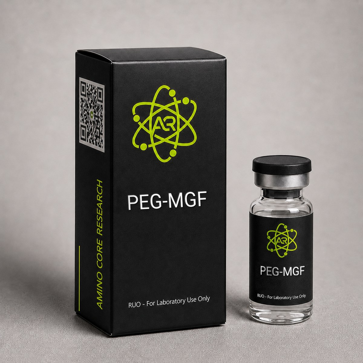
≥99%HPLC Purity
COAIncluded
USAShipped
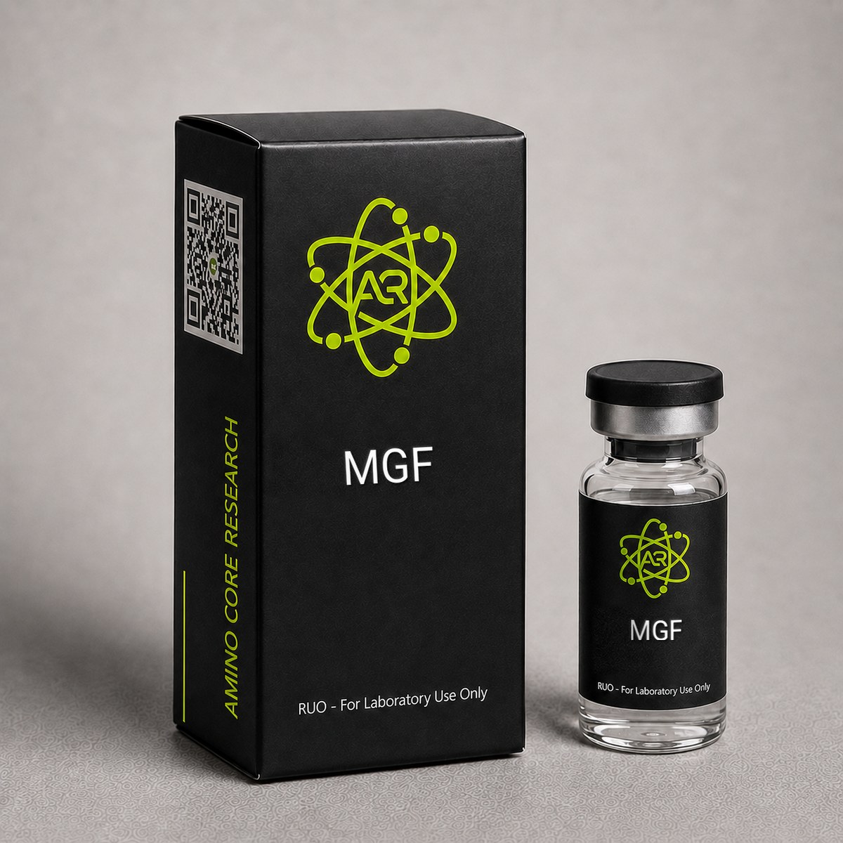
MGF (Mechano Growth Factor) Peptide
Splice variant of IGF-1 expressed in response to mechanical stimulation. 24-amino acid peptide studied for muscle biology research.
Research Reference
This page is a research monograph. MGF (Mechano Growth Factor) is documented here for reference — structure, mechanism and published literature — and is not offered for sale.
Browse compounds available for research purchase · Read the research library
Quick Facts
| SKU | ACR-MGF |
|---|---|
| CAS Number | 62031-54-3 |
| Molecular Formula | C121H200N42O39 |
| Molecular Weight | 2867.14 g/mol |
| Sequence | 24-amino acid IGF-1 splice variant |
| Purity | ≥99% |
| Physical Form | Lyophilized Powder |
| Storage | Store at -20°C |
What is MGF?
Mechano Growth Factor (MGF) is a splice variant of IGF-1 produced in response to mechanical stress on muscle tissue. When the IGF-1 gene is activated by mechanical loading, it undergoes alternative splicing to produce MGF (IGF-1Ec in humans, IGF-1Eb in rodents) rather than the liver-type systemic IGF-1Ea isoform.
The active C-terminal E-domain peptide of MGF consists of 24 amino acids and is responsible for its unique biological activity. Published research has investigated MGF expression in exercised and damaged muscle tissue and its role in satellite cell activation and proliferation.
For laboratory research use only.
Mechanism of Action
Mechano Growth Factor (MGF) is a splice variant of the insulin-like growth factor-1 (IGF-1) gene, specifically the IGF-1Ec isoform in humans (IGF-1Eb in rodents). Unlike systemic IGF-1, which is predominantly produced by the liver under growth hormone stimulation, MGF is expressed locally in skeletal muscle in response to mechanical loading, stretch, and damage. The 24-amino acid C-terminal E-domain peptide (sequence: YQPPSTNKNTKSQRRKGSTFEERK) is the focus of most research, as this region distinguishes MGF from mature IGF-1 and appears to mediate its unique biological actions.[1]
Alternative Splicing and Local Expression
The IGF1 gene undergoes alternative splicing in response to mechanical signals. Inclusion of exon 5 produces the MGF transcript (IGF-1Ec), which encodes a precursor protein with a unique E-peptide. Research suggests that mechanical overload, eccentric contraction, and muscle damage all upregulate MGF mRNA within hours, preceding the slower rise of systemic IGF-1Ea. This temporal pattern positions MGF as an early-response factor in the muscle repair cascade.[2]
Satellite Cell Activation
The most studied action of the synthetic MGF E-peptide is activation of muscle satellite cells (resident myogenic stem cells). In preclinical models, MGF has been shown to recruit quiescent satellite cells into the cell cycle, promoting proliferation prior to terminal differentiation. This is mechanistically distinct from mature IGF-1, which preferentially drives myoblast differentiation and fusion. The dual phases — MGF-mediated proliferation followed by IGF-1Ea-mediated differentiation — represent a coordinated regenerative program.[1]
Receptor Pharmacology
The E-domain peptide does not appear to bind the IGF-1 receptor (IGF-1R) with high affinity. Research suggests MGF acts through a separate, as-yet incompletely characterized receptor or binding site, which may explain its distinct biological profile relative to full-length IGF-1. Downstream signaling has been associated with activation of PI3K/Akt pathways and modulation of MAPK signaling, contributing to anti-apoptotic effects in stressed myocytes and neurons.[3]
Cytoprotective Signaling
In addition to satellite cell effects, the MGF E-peptide has been associated with neuroprotective and cardioprotective actions in preclinical models of ischemia. Research suggests it reduces apoptosis in vulnerable cell populations following hypoxic injury, an effect attributed to upregulation of anti-apoptotic Bcl-2 family proteins and suppression of caspase activation.[3]
Comparison to IGF-1 and IGF-1 LR3
Whereas long-R3 IGF-1 (LR3) is engineered for prolonged systemic IGF-1R activation and broad anabolic signaling, MGF is studied as a short-acting, locally expressed regulator with a specialized role in satellite cell biology. The synthetic MGF peptide has a notably short plasma half-life (minutes), which has driven interest in PEGylated MGF analogs to extend exposure in research settings.[2]
Research & Clinical Studies
Preclinical Study: MGF and Satellite Cell Activation After Muscle Damage
A landmark investigation by Hill and Goldspink (2003) examined the temporal expression of IGF-1 splice variants in rodent skeletal muscle following mechanical overload and damage. The study used a stretch and electrical stimulation protocol on rat tibialis anterior muscle to induce controlled mechanical injury, followed by quantitative analysis of IGF-1Ea and IGF-1Ec (MGF) mRNA expression over a 7-day time course.[1]
Study Design
- Species: adult male Sprague-Dawley rats
- Intervention: passive stretch combined with high-frequency electrical stimulation of TA muscle
- Endpoints: MGF and IGF-1Ea mRNA quantification, satellite cell marker expression (M-cadherin, c-Met)
- Sampling: 0, 6, 24, 48, 72 hours and 7 days post-injury
Key Findings
- MGF mRNA expression increased several-fold within 6 hours post-stimulation, peaking at 24-48 hours
- Mature IGF-1Ea expression rose more slowly, peaking at 72 hours to 7 days, consistent with a downstream differentiation phase
- Satellite cell activation markers M-cadherin and c-Met co-localized with regions of MGF expression
- The temporal sequence supported a model in which MGF drives the proliferative phase of muscle repair, followed by IGF-1Ea-mediated differentiation
This study provided foundational evidence that the IGF-1Ec splice variant is mechanically responsive and that its expression precedes systemic IGF-1 in damaged muscle. The findings have been widely cited as the basis for using synthetic MGF E-peptide in research models of muscle regeneration.[1]
Follow-up Work
Subsequent studies by Goldspink and colleagues demonstrated that age-related decline in MGF responsiveness correlates with reduced satellite cell pools in elderly subjects, suggesting MGF biology is a relevant axis in sarcopenia research. Additional work has been associated with attenuated MGF expression in dystrophic muscle, indicating possible relevance to muscular dystrophy disease models.[2]
[1] Hill M, Goldspink G. Expression and splicing of the insulin-like growth factor gene in rodent muscle is associated with muscle satellite (stem) cell activation following local tissue damage. J Physiol. 2003;549(Pt 2):409-418. PubMed ↗
[2] Goldspink G. Mechanical signals, IGF-I gene splicing, and muscle adaptation. Physiology (Bethesda). 2005;20:232-238. PubMed ↗
Comparative Study: MGF vs IGF-1 on Myoblast Proliferation
One of the foundational comparative studies examining MGF and mature IGF-1 was conducted by Yang and Goldspink (2002), who isolated the unique C-terminal E-domain peptide of MGF and compared its biological activity against full-length IGF-1 in cultured C2C12 myoblasts. This work established a key concept in muscle biology research: MGF and IGF-1, although derived from the same gene, exhibit distinct temporal and functional roles in muscle adaptation.
Study Design:
- Cultured C2C12 murine myoblasts treated with either synthetic MGF E-domain peptide or recombinant mature IGF-1
- Concentrations ranging from 1-100 ng/mL
- Outcome measures: cell proliferation (BrdU incorporation), differentiation markers (myogenin, MHC), and timing of myotube formation
Key Results:
- MGF E-domain peptide stimulated myoblast proliferation without inducing terminal differentiation
- Mature IGF-1 promoted differentiation into myotubes with upregulation of myogenin and myosin heavy chain (MHC)
- MGF maintained cells in a proliferative state — consistent with satellite cell pool expansion
- The two isoforms appeared to act through distinct signaling routes, since MGF activity persisted in cells with IGF-1R knockdown, suggesting a separate receptor or alternative binding mechanism
Context and Significance: This study provided early mechanistic support for the "two-step" hypothesis of muscle adaptation: MGF is expressed first after mechanical loading or damage to expand the satellite cell pool, while mature IGF-1 (IGF-1Ea) is expressed later to drive differentiation and protein synthesis. Subsequent research by Hill and Goldspink (2003) extended these findings in vivo, showing that intramuscular injection of MGF cDNA in rodents produced significant increases in muscle fiber cross-sectional area within 2 weeks — a magnitude exceeding that of IGF-1Ea cDNA injection over the same timeframe.
Comparative work with IGF-1 LR3 (Long R3 IGF-1) shows yet another distinct profile: LR3 has reduced IGFBP binding and a prolonged half-life, predominantly driving IGF-1R-mediated anabolic signaling, whereas MGF appears to act locally and transiently on quiescent satellite cells. Together, these comparative studies position MGF as a research tool uniquely suited to investigating satellite cell biology, regenerative myogenesis, and the early phase of mechanical load adaptation rather than direct hypertrophic signaling.
[1] Yang SY, Goldspink G. Different roles of the IGF-I Ec peptide (MGF) and mature IGF-I in myoblast proliferation and differentiation. FEBS Lett. 2002;522(1-3):156-160. PubMed ↗
[2] Hill M, Goldspink G. Expression and splicing of the insulin-like growth factor gene in rodent muscle is associated with muscle satellite (stem) cell activation following local tissue damage. J Physiol. 2003;549(Pt 2):409-418. PubMed ↗
Chemical & Physical Properties
The following properties characterize the synthetic MGF E-domain peptide as supplied by AminoCore Research for in vitro and preclinical investigation.
| Full Name | Mechano Growth Factor (E-domain peptide) |
|---|---|
| Synonyms | MGF, IGF-1Ec E-peptide, IGF-1 Ec |
| Molecular Formula | C121H200N42O39 |
| Molecular Weight | 2,867.14 g/mol |
| CAS Number | 62031-54-3 |
| Sequence | Tyr-Gln-Pro-Pro-Ser-Thr-Asn-Lys-Asn-Thr-Lys-Ser-Gln-Arg-Arg-Lys-Gly-Ser-Thr-Phe-Glu-Glu-Arg-Lys (YQPPSTNKNTKSQRRKGSTFEERK) |
| Amino Acid Count | 24 |
| Origin / Developer | Identified by Goldspink and colleagues (University of London) as a mechano-sensitive splice variant of IGF-1 |
| Key Modifications | Synthetic C-terminal E-peptide derived from the IGF-1Ec exon 5 splice variant; unmodified backbone |
| Source Gene | IGF1 (alternative splicing including exon 5) |
| Physical Form | Lyophilized white powder |
| Solubility | Soluble in bacteriostatic or sterile water; soluble in dilute acetic acid for stock preparation |
| Stability | Short plasma half-life in solution (minutes); lyophilized form stable for extended periods at -20°C |
| Purity | ≥99% by HPLC |
| Appearance Post-Reconstitution | Clear, colorless solution |
Notes on Identity
The peptide marketed and studied as "MGF" in research settings refers specifically to the 24-residue C-terminal E-domain of the IGF-1Ec splice variant, not the full-length pro-IGF-1Ec protein. Investigators should note this distinction when comparing literature, as some studies refer to the full pro-peptide while research-grade synthetic MGF reflects only the unique E-domain sequence. The peptide is highly basic (multiple lysine and arginine residues), contributing to its short circulating half-life and rapid renal clearance in preclinical models.
Handling & Reconstitution Guidelines
MGF (Mechano Growth Factor) is supplied as a sterile lyophilized powder. As a 24-amino acid peptide containing multiple polar residues and a free N-terminus, it requires careful reconstitution to preserve structural integrity and biological activity in research applications. Proper handling minimizes peptide degradation, aggregation, and loss of activity due to surface adsorption.
Recommended Reconstitution Protocol:
- Equilibrate the vial to room temperature (15-20 minutes) before opening to prevent moisture condensation on the lyophilized powder.
- Briefly centrifuge the vial (if equipment is available) to ensure the lyophilized cake is at the bottom prior to opening.
- Select reconstitution solvent: bacteriostatic water (0.9% benzyl alcohol) is the standard for research reconstitution. Sterile water or 0.9% sodium chloride may also be used for short-term studies.
- Calculate concentration: for a 2 mg vial reconstituted with 1 mL of bacteriostatic water, the final concentration is 2 mg/mL (2000 µg/mL). Adjust solvent volume to achieve the desired working concentration.
- Inject solvent slowly down the inner wall of the vial — do not direct the stream onto the lyophilized powder, which can cause foaming and denaturation.
- Swirl gently until fully dissolved. Do not shake or vortex — mechanical agitation can shear peptide bonds and induce aggregation.
- Inspect the solution: it should appear clear and colorless. Any visible particulates indicate compromised material that should not be used.
Compound-Specific Notes:
- MGF has a notably short half-life in solution — research data suggest functional activity declines within minutes to hours after reconstitution in physiological buffers, due to rapid proteolytic cleavage of the E-domain peptide.
- For in vitro experiments, reconstituted aliquots should be used immediately or stored at -20°C in single-use portions to avoid freeze-thaw cycles.
- Use low-binding polypropylene tubes for storage and dilution. Glass and high-binding plastics can cause significant peptide loss via surface adsorption.
- Avoid extended exposure to light or temperatures above 8°C once reconstituted.
All handling should be performed using appropriate personal protective equipment (PPE) including gloves and lab coat. MGF is for laboratory research use only and is not intended for human or veterinary administration.
Storage & Stability Information
Proper storage is critical for maintaining the structural integrity and biological activity of MGF (Mechano Growth Factor). As a 24-amino acid splice-variant peptide, MGF is sensitive to temperature fluctuations, moisture, and repeated freeze-thaw cycles. The following guidelines reflect standard practice for research-grade peptides of this class.
Lyophilized Powder Storage:
- Long-term (recommended): Store at -20°C or below in a frost-free freezer. Under these conditions, lyophilized MGF retains stability for 24 months or longer when protected from moisture and light.
- Short-term (up to 30 days): Store at 2-8°C in a sealed, desiccated container.
- Transit/shipping: Brief exposure to room temperature (≤7 days) during shipping does not significantly compromise lyophilized peptide quality.
- Keep the vial sealed and protected from light. Do not open until ready to reconstitute.
Reconstituted Solution Storage:
- 2-8°C: Reconstituted MGF should be used within 5-7 days when stored refrigerated in bacteriostatic water.
- -20°C (aliquoted): Single-use aliquots in low-binding polypropylene tubes can be stored for up to 30 days with minimal activity loss.
- Avoid repeated freeze-thaw cycles — each cycle can reduce active peptide concentration by 5-15% due to aggregation and proteolytic fragmentation.
Compound-Specific Stability Notes:
- MGF's C-terminal E-domain is highly susceptible to proteolytic cleavage in aqueous solution, particularly at physiological pH. This is the basis for its naturally short half-life in vivo and informs why fresh reconstitution is preferred for time-sensitive experiments.
- The peptide contains no cysteine residues, so disulfide-mediated aggregation is not a concern; however, the high content of polar and charged residues makes it prone to surface adsorption on glass and untreated plastic.
- Methionine and tryptophan residues are sensitive to oxidation — minimize exposure to atmospheric oxygen by keeping vials capped and storing under inert conditions when possible for extended studies.
- Avoid storage in standard household freezers with frequent door openings; temperature cycling accelerates degradation.
Adherence to these storage parameters supports reproducibility across in vitro and ex vivo MGF research applications.
Frequently Asked Questions
What is MGF?
MGF (Mechano Growth Factor) is a splice variant of IGF-1 produced by mechanical muscle loading. The active 24-amino acid C-terminal peptide has been studied for satellite cell activation. It differs from systemic IGF-1 in tissue specificity. For research use only.
How does MGF differ from IGF-1 and IGF-1 LR3?
MGF (Mechano Growth Factor) is a splice variant of the IGF-1 gene — specifically the IGF-1Ec isoform — that is expressed locally in skeletal muscle in response to mechanical loading. The research peptide consists of the unique 24-amino acid C-terminal E-domain that distinguishes MGF from mature IGF-1. Functionally, research suggests MGF preferentially activates satellite cell proliferation, whereas mature IGF-1 (and the engineered long-acting analog IGF-1 LR3) drives myoblast differentiation and broad anabolic signaling through the IGF-1 receptor. MGF also has a much shorter plasma half-life (minutes versus hours for LR3) and appears to act through a binding site distinct from IGF-1R.
What is the molecular weight and CAS number of MGF?
The synthetic MGF E-peptide has a molecular weight of 2,867.14 g/mol, a molecular formula of C121H200N42O39, and CAS number 62031-54-3. The 24-amino acid sequence is YQPPSTNKNTKSQRRKGSTFEERK, corresponding to the C-terminal E-domain of the human IGF-1Ec splice variant. AminoCore Research supplies MGF as a lyophilized powder at ≥99% HPLC purity for in vitro and preclinical research use only.
How should MGF be stored and reconstituted?
Lyophilized MGF should be stored at -20°C for long-term stability, with short-term storage at 2-8°C acceptable for up to a few weeks. For reconstitution, bacteriostatic water or sterile water is typically used; the vial should be swirled gently rather than shaken to avoid peptide degradation. Once reconstituted, MGF has limited stability in solution and should be kept at 2-8°C and used within several days, or aliquoted and stored at -20°C to minimize freeze-thaw cycles. The peptide is highly basic and sensitive to repeated thawing.
Does MGF act through the IGF-1 receptor?
Research suggests that the synthetic 24-amino acid MGF E-peptide does not bind the IGF-1 receptor (IGF-1R) with high affinity, which distinguishes it from mature IGF-1 and engineered analogs like LR3. Instead, the E-domain appears to act through a separate, incompletely characterized binding site or receptor that mediates satellite cell activation and cytoprotective signaling. Downstream pathways have been associated with PI3K/Akt and MAPK activation, contributing to anti-apoptotic effects. This pharmacological separation is a key reason MGF is studied as a distinct entity from IGF-1 in muscle biology research.
What sizes of MGF are available from AminoCore Research?
MGF (Mechano Growth Factor) is typically offered in 2 mg and 5 mg lyophilized vials at ≥99% HPLC purity. Each vial is supplied with a Certificate of Analysis (COA) documenting purity, mass spectrometry confirmation of molecular weight (2867.14 g/mol), and lot identification. All material is sold strictly for laboratory research use and is not intended for human consumption, veterinary use, or therapeutic application. Researchers requiring custom quantities or bulk pricing for institutional studies should contact AminoCore Research directly to discuss availability.
Why is MGF said to have such a short half-life in solution?
MGF is a 24-amino acid synthetic peptide corresponding to the unique C-terminal E-domain of the IGF-1Ec splice variant. In aqueous solution and biological fluids, this small peptide is rapidly cleaved by endogenous peptidases, with research literature reporting functional half-lives on the order of minutes after administration in preclinical models. This short half-life reflects MGF's natural biological role as a transient, local autocrine/paracrine signal expressed by mechanically stressed muscle fibers. For research applications, this property means MGF is typically reconstituted immediately before use and is best suited to studies examining acute satellite cell activation rather than sustained anabolic signaling.
Does MGF act on the same receptor as IGF-1?
Current research evidence suggests MGF does NOT primarily signal through the classical IGF-1 receptor (IGF-1R). Studies by Yang and Goldspink (2002) and subsequent work demonstrated that the synthetic MGF E-domain peptide retains biological activity in cells where IGF-1R is knocked down or blocked, indicating a distinct molecular target. The exact MGF receptor has not been definitively identified, though candidate binding partners and alternative signaling routes involving ERK/MAPK pathways have been proposed. This receptor independence is one of the most scientifically interesting features of MGF and is an active area of muscle biology research.
How does MGF compare to IGF-1 LR3 for muscle research applications?
MGF and IGF-1 LR3 (Long R3 IGF-1) are distinct research tools targeting different aspects of muscle biology. IGF-1 LR3 is a modified mature IGF-1 analog with reduced IGFBP binding and a half-life of approximately 20-30 hours, producing sustained activation of the IGF-1 receptor and downstream PI3K/Akt/mTOR signaling — well suited to studying chronic anabolic and hypertrophic pathways. MGF, by contrast, is the E-domain peptide of the IGF-1Ec splice variant with a very short half-life, appearing to act through non-IGF-1R mechanisms to activate quiescent satellite cells and promote myoblast proliferation. Researchers typically use LR3 for sustained anabolic signaling studies and MGF for satellite cell, regeneration, and acute mechanical load response models.
For laboratory and research use only. This product is not for human or veterinary use. It is not a drug, supplement or food.
This product is not FDA approved for any indication, and is not intended to diagnose, treat, cure, or prevent any disease. All product information is derived from published preclinical research and does not constitute medical advice or claims.