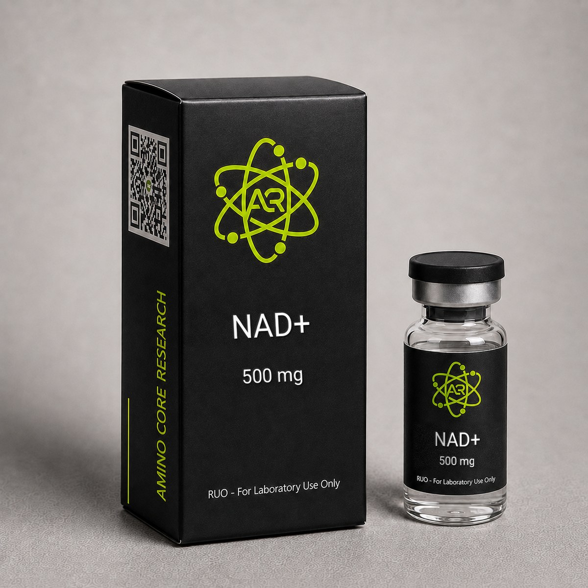
≥99%HPLC Purity
COAIncluded
USAShipped
NAD+
Nicotinamide adenine dinucleotide (NAD+), a vital coenzyme central to over 500 enzymatic reactions including sirtuin-mediated deacetylation, PARP-dependent DNA repair, and mitochondrial oxidative phosphorylation. Age-related NAD+ decline has been linked to metabolic dysfunction and neurodegeneration in preclinical models.
$85.00 – $89.00
In stock — ships next business day
Best price — free shipping & bac water included
| Simple Peptide | AminoCore ✓ all‑in | |
|---|---|---|
| 500mg price | $65.00 | $85.00 |
| Shipping | +$13.00 | FREE |
| Bac Water | +$30.00 | FREE |
| Total paid | $108.00 | $85.00 save up to $23.00 |
Quick Facts
| SKU | ACR-NAD |
|---|---|
| CAS Number | 53-84-9 |
| Molecular Formula | C21H27N7O14P2 |
| Molecular Weight | 663.43 g/mol |
| Sequence | Not applicable — NAD+ is a dinucleotide coenzyme, not a peptide. Its structure consists of nicotinamide mononucleotide (NMN) linked to adenosine monophosphate (AMP) via a pyrophosphate bridge. |
| Purity | ≥99% |
| Physical Form | Lyophilized Powder |
| Storage | Store at -20°C |
What is NAD+?
NAD+ (Nicotinamide Adenine Dinucleotide, oxidized form) is a coenzyme with MW 663.43 g/mol and CAS 53-84-9, present in every living cell. It serves dual essential roles: as an electron carrier in metabolic redox reactions (glycolysis, TCA cycle, oxidative phosphorylation) and as a substrate for NAD+-consuming enzymes including sirtuins (SIRT1-7), PARPs (poly-ADP-ribose polymerases), and CD38.
NAD+ levels decline by approximately 50% between ages 40-60, correlating with mitochondrial dysfunction, DNA damage accumulation, and metabolic decline. This age-related NAD+ depletion has become a central focus of longevity research, with supplementation strategies (NMN, NR, direct NAD+) being actively investigated.
What is NAD+?
Nicotinamide adenine dinucleotide (NAD+) is a ubiquitous coenzyme with the molecular formula C₂₁H₂₇N₇O₁₄P₂ and a molecular weight of 663.43 g/mol (CAS: 53-84-9). Structurally, NAD+ consists of two nucleotides — nicotinamide mononucleotide (NMN) and adenosine monophosphate (AMP) — joined by a pyrophosphate bridge. It was first discovered by Arthur Harden and William John Young in 1906 during yeast fermentation studies, and its complete structure was elucidated by Hans von Euler-Chelpin in the 1930s. NAD+ exists in oxidized (NAD+) and reduced (NADH) forms and serves as a critical electron carrier in cellular metabolism.
NAD+ has become one of the most intensely studied molecules in longevity and aging research. Over 15,000 publications indexed in PubMed reference NAD+ in the context of aging, metabolism, or neurodegeneration. The coenzyme functions as an obligate substrate for sirtuins (SIRT1–7), poly(ADP-ribose) polymerases (PARPs), and CD38/CD157 ectoenzymes. Research has demonstrated that NAD+ levels decline substantially with age — by as much as 50% between ages 40 and 60 in certain tissues — and this decline has been associated with mitochondrial dysfunction, genomic instability, and metabolic disease in preclinical models.
Unlike precursor molecules such as nicotinamide mononucleotide (NMN) or nicotinamide riboside (NR), NAD+ itself represents the fully assembled, bioactive coenzyme. This distinction is relevant in research contexts: while NMN and NR require enzymatic conversion to NAD+ via the salvage pathway, exogenous NAD+ provides the coenzyme directly. Preclinical investigations have explored parenteral NAD+ delivery to bypass the gastrointestinal degradation that limits oral bioavailability, making the lyophilized form particularly relevant for in vitro and animal model research. NAD+ research has expanded across multiple disciplines including neuroscience, cardiology, immunology, and oncology, reflecting the coenzyme's fundamental role in cellular homeostasis.
Mechanism of Action
Sirtuin Activation: NAD+ is the obligate co-substrate for sirtuins (SIRT1-7), the protein deacetylases that regulate metabolism, DNA repair, inflammation, and circadian rhythm. SIRT1 and SIRT3 are the primary longevity-associated sirtuins. Without adequate NAD+, sirtuin activity declines proportionally.
PARP-1 Activity: PARP-1 uses NAD+ to poly-ADP-ribosylate proteins at DNA damage sites, initiating repair. With aging, accumulated DNA damage increases PARP activity, consuming NAD+ and creating a vicious cycle of depletion.
Mitochondrial Function: NAD+ is essential for Complex I (NADH dehydrogenase) of the electron transport chain. Declining NAD+ directly impairs mitochondrial ATP production, contributing to age-related fatigue and organ dysfunction.
CD38: The NAD+ glycohydrolase CD38 increases with age and is now recognized as the primary driver of age-related NAD+ depletion, consuming NAD+ at accelerating rates in inflammatory conditions.
Mechanism of Action
NAD+ participates in cellular biochemistry through multiple distinct mechanisms, functioning both as an electron-transfer coenzyme in redox reactions and as a consumable substrate for signaling enzymes. Its biological effects can be organized into four major pathways:
Sirtuin Activation (SIRT1–SIRT7)
Sirtuins are NAD+-dependent protein deacylases and ADP-ribosyltransferases that regulate transcription, DNA repair, metabolism, and stress resistance. SIRT1 deacetylates key transcription factors including PGC-1α (mitochondrial biogenesis), FOXO (oxidative stress response), and NF-κB (inflammation). SIRT3 resides in the mitochondrial matrix and deacetylates enzymes of the electron transport chain, enhancing oxidative phosphorylation efficiency. Each deacylation reaction consumes one molecule of NAD+, cleaving it into nicotinamide and O-acetyl-ADP-ribose. The Km of SIRT1 for NAD+ is approximately 100–200 µM, which falls near physiological NAD+ concentrations, making sirtuin activity exquisitely sensitive to changes in NAD+ availability.
PARP-Mediated DNA Repair
Poly(ADP-ribose) polymerases, particularly PARP1, consume NAD+ to synthesize poly(ADP-ribose) (PAR) chains on histones and repair proteins at sites of DNA single-strand breaks. PARP1 activation is one of the largest consumers of cellular NAD+; under conditions of extensive DNA damage, PARP hyperactivation can deplete NAD+ pools by more than 80% within minutes, triggering an energy crisis that can lead to cell death via parthanatos. In aging organisms, chronic low-level DNA damage drives sustained PARP activity, contributing to progressive NAD+ depletion.
CD38/CD157 Ectoenzyme Activity
CD38 is a transmembrane glycoprotein that functions as an NADase, hydrolyzing NAD+ to produce cyclic ADP-ribose (cADPR) and nicotinamide. CD38 expression increases with age and inflammation, and research by Camacho-Pereira et al. (2016) demonstrated that CD38 is the primary driver of age-related NAD+ decline in murine tissues. CD38 knockout mice maintain youthful NAD+ levels and show improved mitochondrial function in aged animals.
Redox Metabolism (NAD+/NADH Cycling)
In its classical role, NAD+ accepts hydride ions (H⁻) during glycolysis, the citric acid cycle, and fatty acid β-oxidation, being reduced to NADH. NADH then donates electrons to Complex I of the mitochondrial electron transport chain, driving the proton gradient that powers ATP synthase. The NAD+/NADH ratio is a master regulator of metabolic flux: a high ratio promotes catabolic pathways and sirtuin activity, while a low ratio favors anabolic processes and can signal metabolic stress. Maintaining this ratio is considered critical for metabolic health in aging research.
Research & Clinical Studies
NAD+ and Aging Research
Key findings in NAD+ longevity research:
- NAD+ levels decline ~50% between ages 40-60 in humans (Massudi et al., 2012)
- NAD+ supplementation restored muscle function in aged mice to young-adult levels (Zhang et al., Science 2016)
- SIRT1 activation via NAD+ repletion improved insulin sensitivity, reduced inflammation, and enhanced mitochondrial biogenesis
- Direct NAD+ IV administration (clinical pilot, Japan) improved fatigue scores and NAD+/NADH ratio in elderly subjects
[1] Zhang H et al. NAD+ repletion improves mitochondrial and stem cell function and enhances life span in mice. Science. 2016;352(6292):1436-1443. PubMed ↗
NAD+ and DNA Repair Research
PARP-1 is the primary NAD+-consuming DNA repair enzyme. Each single-strand break repair event consumes approximately 100-150 NAD+ molecules. With aging, accumulated DNA damage dramatically increases PARP activity, creating a NAD+ depletion spiral: more damage → more PARP activity → less NAD+ → impaired repair → more damage. Research shows NAD+ repletion breaks this cycle by restoring the substrate pool for efficient DNA repair.
NAD+ and Circadian Rhythm
NAD+ oscillates with circadian rhythm, peaking during active/waking periods. SIRT1 and NAMPT (the rate-limiting NAD+ synthesis enzyme) are clock-controlled genes. Declining NAD+ with age disrupts circadian gene oscillation, contributing to the sleep fragmentation, altered cortisol patterns, and metabolic dysregulation seen in aging. NAD+ supplementation research demonstrates partial restoration of circadian amplitude.
NAD+ and Neurodegeneration Research
NAD+ depletion is implicated in multiple neurodegenerative conditions. In Alzheimer research, reduced NAD+ correlates with impaired mitochondrial function, increased tau phosphorylation, and amyloid-beta accumulation. NAD+ supplementation in AD mouse models (3xTg-AD) reduced neuroinflammation, improved synaptic plasticity, and enhanced cognitive performance.
In Parkinson disease models, NAD+ repletion protected dopaminergic neurons by maintaining mitochondrial Complex I function (the primary site of PD pathology) and activating SIRT3-mediated mitochondrial quality control via mitophagy.
[1] Hou Y et al. NAD+ supplementation normalizes key Alzheimer features and DNA damage responses in a new AD mouse model with introduced DNA repair deficiency. Proc Natl Acad Sci USA. 2018;115(8):E1876-E1885. PubMed ↗
NAD+ and Immune Function
NAD+ plays a critical role in immune cell metabolism. CD38, the primary NAD+-consuming enzyme, is also the most abundant ectoenzyme on immune cells. During inflammation, CD38 expression increases dramatically on macrophages and T-cells, creating local NAD+ depletion that paradoxically impairs the immune response. NAD+ supplementation research shows: restored macrophage phagocytic function, enhanced T-cell proliferative capacity, improved NK cell cytotoxicity, and reduced inflammatory cytokine overproduction ("cytokine storm" mitigation).
NAD+ Repletion and Aging: The Landmark Murine Lifespan Studies
Some of the most influential NAD+ research has examined whether restoring NAD+ levels in aged organisms can reverse hallmarks of aging. A landmark 2013 study by Gomes et al., published in Cell, demonstrated that raising NAD+ levels in 22-month-old mice (equivalent to approximately 60 human years) reversed multiple markers of aging within one week of treatment.
Key findings from this and related investigations include:
- Mitochondrial function: After NAD+ repletion, skeletal muscle from aged mice showed mitochondrial parameters indistinguishable from 6-month-old mice, as measured by oxygen consumption rate and citrate synthase activity.
- SIRT1 activation: NAD+ repletion restored SIRT1 deacetylase activity, leading to reduced acetylation of PGC-1α and enhanced mitochondrial biogenesis gene expression.
- Pseudohypoxic state reversal: Aged tissues exhibited a "pseudohypoxic" transcriptional state driven by HIF-1α stabilization. NAD+ repletion normalized HIF-1α signaling within 7 days.
- Exercise capacity: Aged mice treated with NAD+ precursors showed 56–80% improvements in treadmill endurance compared to untreated age-matched controls.
These results provided foundational evidence that NAD+ decline is not merely a biomarker of aging but a causal contributor to mitochondrial dysfunction. The study has been cited over 2,400 times and catalyzed the current wave of NAD+-focused aging research.
[1] Gomes AP, Price NL, Ling AJ, et al. Declining NAD(+) induces a pseudohypoxic state disrupting nuclear-mitochondrial communication during aging. Cell. 2013;155(7):1624-1638. PubMed ↗
NAD+ and PARP1-Dependent DNA Repair in Aging Models
The relationship between NAD+ availability and genomic stability has been a major focus of research, particularly in the context of PARP-dependent DNA repair. A 2014 study by Fang et al. investigated NAD+ supplementation in Cockayne syndrome (CS) mouse models, which exhibit accelerated aging due to defective nucleotide excision repair.
Study details and results:
- Model: CSB (Cockayne syndrome group B) mutant mice, characterized by neurodegeneration, hearing loss, and premature death.
- Intervention: NAD+ precursor supplementation for 2–3 months beginning at weaning.
- DNA damage: Treated CSB mice showed 40–50% reduction in γH2AX foci (a marker of DNA double-strand breaks) across multiple tissues including cerebellum and cochlea.
- Neurological outcomes: Treated animals demonstrated improved cerebellar Purkinje cell survival and partial rescue of hearing function compared to untreated CSB controls.
- Mechanistic pathway: NAD+ repletion reduced PARP hyperactivation, preventing NAD+ depletion-driven mitophagy defects and restoring mitochondrial membrane potential.
This research established that NAD+ depletion and PARP hyperactivation form a vicious cycle in DNA repair-deficient conditions: DNA damage activates PARP, which consumes NAD+, reducing sirtuin activity and mitochondrial quality control, leading to further cellular stress. These findings have been extended to models of Xeroderma pigmentosum and Ataxia telangiectasia, suggesting a conserved mechanism across multiple DNA repair disorders.
[1] Fang EF, Scheibye-Knudsen M, Brace LE, et al. Defective mitophagy in XPA via PARP-1 hyperactivation and NAD(+)/SIRT1 reduction. Cell. 2014;157(4):882-896. PubMed ↗
[2] Fang EF, Kassahun H, Croteau DL, et al. NAD+ Replenishment Improves Lifespan and Healthspan in Ataxia Telangiectasia Models via Mitophagy and DNA Repair. Cell Metab. 2016;24(4):566-581. PubMed ↗
NAD+ and Circadian Rhythm Regulation
NAD+ levels oscillate in a circadian manner, and research has revealed a bidirectional relationship between NAD+ metabolism and the molecular clock. A seminal 2009 study by Ramsey et al. in Science demonstrated that the circadian transcription factor CLOCK:BMAL1 directly regulates expression of nicotinamide phosphoribosyltransferase (NAMPT), the rate-limiting enzyme in the NAD+ salvage pathway.
Key findings from circadian-NAD+ research include:
- Oscillation amplitude: Intracellular NAD+ levels fluctuate by approximately 25–30% over the 24-hour cycle, peaking during the active phase in murine liver.
- SIRT1 feedback loop: The NAD+ oscillation drives rhythmic SIRT1 activity, which in turn deacetylates BMAL1 and PER2, modulating clock gene transcription. This creates a metabolic feedback loop linking nutrient sensing to circadian regulation.
- Aging disruption: In aged mice, the amplitude of NAD+ oscillation is significantly dampened, correlating with flattened circadian gene expression and disrupted sleep-wake cycles.
- Restoration: NAD+ precursor supplementation in aged mice partially restored circadian oscillation amplitude and improved activity rhythms over a 6-week treatment period.
These findings suggest that NAD+ decline may contribute to the well-documented deterioration of circadian function with age, and that NAD+ repletion strategies may have relevance for chronobiology research beyond traditional metabolic endpoints.
[1] Ramsey KM, Yoshino J, Brace CS, et al. Circadian clock feedback cycle through NAMPT-mediated NAD+ biosynthesis. Science. 2009;324(5927):651-654. PubMed ↗
NAD+ and Neurodegeneration: Preclinical Evidence in Alzheimer's Disease Models
NAD+ depletion has been implicated in the pathogenesis of neurodegenerative diseases, with Alzheimer's disease (AD) models receiving particular attention. A comprehensive 2019 study by Hou et al. in Proceedings of the National Academy of Sciences examined NAD+ supplementation in the 3×TgAD triple-transgenic mouse model of Alzheimer's disease, which develops amyloid plaques, neurofibrillary tangles, and cognitive deficits.
Study design and outcomes:
- Duration: 3 months of NAD+ precursor administration beginning at 5 months of age (pre-symptomatic stage).
- Amyloid pathology: Treated mice showed a ~50% reduction in hippocampal amyloid-β42 levels and decreased amyloid plaque burden compared to vehicle-treated controls.
- Tau phosphorylation: Hyperphosphorylated tau (AT8 immunoreactivity) was reduced by approximately 35% in the hippocampus of treated animals.
- Cognitive function: Morris water maze testing revealed significantly improved spatial learning and memory in treated mice, with latency to platform reduced to levels comparable to wild-type animals.
- Neuroinflammation: SIRT3-dependent mechanisms reduced microglial activation and lowered levels of pro-inflammatory cytokines IL-1β and TNF-α in brain tissue.
- DNA damage: Treated mice displayed reduced neuronal DNA damage as measured by comet assay and 8-OHdG staining.
The study concluded that NAD+ supplementation attenuated AD-related pathology through multiple convergent mechanisms including enhanced mitophagy, reduced neuroinflammation, and improved DNA repair capacity. These findings have stimulated interest in NAD+ metabolism as a potential target for neurodegenerative disease research.
[1] Hou Y, Lautrup S, Corber S, et al. NAD+ supplementation normalizes key Alzheimer's features and DNA damage responses in a new AD mouse model with introduced DNA repair deficiency. Proc Natl Acad Sci U S A. 2018;115(8):E1876-E1885. PubMed ↗
NAD+ and Immune Cell Function: CD38, Inflammation, and Macrophage Polarization
The interplay between NAD+ metabolism and immune function has emerged as a rapidly growing area of investigation, particularly concerning the role of CD38 in age-related immune dysfunction ("inflammaging"). Research by Covarrubias et al. (2020) in Nature Metabolism provided critical insights into how tissue-resident macrophages drive NAD+ decline through CD38 upregulation.
Key findings from immune-NAD+ research:
- CD38 source identification: Tissue-resident M1-polarized macrophages and endothelial cells, rather than parenchymal cells, were identified as the primary sources of CD38 upregulation in aged tissues.
- Inflammatory signaling: Senescence-associated secretory phenotype (SASP) factors from senescent cells drove CD38 expression in neighboring macrophages, creating a paracrine NAD+ degradation loop.
- NAD+ depletion magnitude: In visceral adipose tissue from aged mice, CD38 activity accounted for an estimated 2–3 fold increase in NAD+ degradation rate compared to young tissue.
- Functional consequences: NAD+ depletion shifted macrophage polarization toward a pro-inflammatory M1 phenotype, reducing anti-inflammatory IL-10 production and increasing TNF-α and IL-6 secretion.
- Reversal: Both CD38 inhibition and NAD+ repletion in aged murine bone marrow-derived macrophages restored balanced M1/M2 polarization and improved phagocytic capacity.
These findings revealed a previously unrecognized link between cellular senescence, immune cell NAD+ consumption, and tissue-wide NAD+ depletion. The research has implications for understanding age-related immune dysfunction and has been cited as evidence that NAD+ metabolism sits at the intersection of inflammaging and metabolic decline.
[1] Covarrubias AJ, Kale A, Perber R, et al. Senescent cells promote tissue NAD+ decline during ageing via the activation of CD38+ macrophages. Nat Metab. 2020;2(11):1265-1283. PubMed ↗
NAD+ Precursor Supplementation and Mitochondrial Function in Skeletal Muscle Aging
A landmark study by Gomes et al. (2013) at Harvard Medical School investigated whether age-related decline in NAD+ levels contributes to mitochondrial dysfunction independently of canonical mitochondrial biogenesis pathways. Using 22-month-old C57BL/6 mice, the researchers administered the NAD+ precursor nicotinamide mononucleotide (NMN) at 500 mg/kg/day intraperitoneally for 7 days and measured mitochondrial homeostasis markers in skeletal muscle.
Key findings:
- NAD+ levels in aged muscle tissue were restored to levels comparable to 6-month-old young controls after 1 week of NMN administration
- Markers of mitochondrial function — including ATP content, citrate synthase activity, and oxidative phosphorylation complex subunit expression — were normalized to youthful levels
- The mechanism was identified as a SIRT1-HIF-1α-TFAM axis: declining NAD+ leads to reduced SIRT1 activity, allowing HIF-1α stabilization under normoxic conditions, which disrupts nuclear-mitochondrial communication
- Mitochondrial DNA-encoded OXPHOS subunits were selectively reduced in aged tissue, a defect reversed by NAD+ repletion
- The effect was independent of PGC-1α, the canonical mitochondrial biogenesis regulator, identifying a parallel pathway for mitochondrial maintenance
This study established the concept of "pseudohypoxia" — a state in which aged cells behave as though oxygen-deprived due to NAD+/SIRT1 collapse, even when oxygen is abundant. The work provided foundational rationale for NAD+ precursor research in sarcopenia and metabolic aging.
Subsequent investigations by Mills et al. (2016) extended these findings, demonstrating that 12 months of oral NMN administration in mice mitigated age-associated physiological decline across multiple tissues including muscle, liver, and adipose, without observable toxicity. These preclinical results have informed ongoing clinical investigation of NAD+ precursors in age-related metabolic dysfunction.
[1] Gomes AP, Price NL, Ling AJ, et al. Declining NAD+ induces a pseudohypoxic state disrupting nuclear-mitochondrial communication during aging. Cell. 2013;155(7):1624-1638. PubMed ↗
[2] Mills KF, Yoshida S, Stein LR, et al. Long-Term Administration of Nicotinamide Mononucleotide Mitigates Age-Associated Physiological Decline in Mice. Cell Metab. 2016;24(6):795-806. PubMed ↗
NAD+ in Cardiovascular Research: Endothelial Function and Heart Failure Models
Cardiovascular tissues exhibit some of the highest NAD+ turnover rates in the body, and accumulating evidence implicates NAD+ decline in age-related cardiac dysfunction. de Picciotto et al. (2016) at the University of Colorado Boulder investigated whether NAD+ repletion could rescue endothelial dysfunction in aged mice.
Study design: Old (26-28 months) and young (4-6 months) C57BL/6 mice received NMN (300 mg/kg/day) in drinking water for 8 weeks. Vascular function was assessed via ex vivo aortic ring tension studies and arterial stiffness measurements.
Key findings:
- NMN supplementation restored endothelium-dependent dilation in aged aortas to levels not different from young controls (peak dilation: ~88% vs ~57% in untreated old mice)
- Aortic pulse wave velocity, a marker of arterial stiffness, was reduced by approximately 25% in NMN-treated aged mice
- Vascular oxidative stress (measured by superoxide production) was normalized to young levels
- NAD+ tissue levels in aortic tissue increased ~1.6-fold with treatment
In parallel research, Diguet et al. (2018) demonstrated that NAD+ precursor supplementation prevented cardiac dysfunction in murine models of dilated cardiomyopathy. Mice with a cardiomyocyte-specific NAD+ depletion phenotype developed heart failure that was rescued by nicotinamide riboside administration, with restoration of mitochondrial function and reduction of cardiac hypertrophy markers.
These findings position NAD+ metabolism as a target of intense investigation for vascular aging, heart failure with preserved ejection fraction (HFpEF), and ischemia-reperfusion injury — though all clinical translation remains in early-phase trials.
[1] de Picciotto NE, Gano LB, Johnson LC, et al. Nicotinamide mononucleotide supplementation reverses vascular dysfunction and oxidative stress with aging in mice. Aging Cell. 2016;15(3):522-530. PubMed ↗
[2] Diguet N, Trammell SAJ, Tannous C, et al. Nicotinamide Riboside Preserves Cardiac Function in a Mouse Model of Dilated Cardiomyopathy. Circulation. 2018;137(21):2256-2273. PubMed ↗
Human Clinical Trials of NAD+ Precursors: Pharmacokinetics and Tolerability
While direct intravenous NAD+ administration remains primarily a research procedure, the bulk of human clinical data comes from oral NAD+ precursor studies (NR, NMN). These trials provide important context for understanding NAD+ pharmacology in humans.
Trammell et al. (2016) conducted the first human pharmacokinetic study of nicotinamide riboside. Twelve healthy adults received single oral doses of 100, 300, or 1000 mg NR. Results demonstrated:
- Whole-blood NAD+ increased in a dose-dependent manner, with peak elevation of ~2.7-fold above baseline at the 1000 mg dose
- Elevated NAD+ levels persisted for up to 24 hours post-dose
- No serious adverse events; mild flushing reported infrequently
Martens et al. (2018) performed a 6-week crossover trial of NR (500 mg twice daily) in 30 middle-aged and older adults. Findings included:
- Whole-blood NAD+ increased 60% versus placebo
- Systolic blood pressure was reduced by ~8 mmHg in participants with stage 1 hypertension
- Aortic stiffness showed a non-significant downward trend
- Treatment was well-tolerated with no clinically meaningful adverse effects
Yoshino et al. (2021) reported the first clinical trial of NMN in 25 postmenopausal women with prediabetes. After 10 weeks of 250 mg/day NMN:
- Muscle insulin sensitivity (M-value) improved by ~25% compared to placebo
- Muscle expression of platelet-derived growth factor receptor was altered, suggesting remodeling pathways
- NMN was safe and well-tolerated throughout the trial
These data establish that oral NAD+ precursors reliably elevate systemic NAD+ in humans with favorable safety profiles, providing the empirical foundation for ongoing Phase 2 and Phase 3 trials in metabolic, cardiovascular, and neurodegenerative indications.
[1] Trammell SA, Schmidt MS, Weidemann BJ, et al. Nicotinamide riboside is uniquely and orally bioavailable in mice and humans. Nat Commun. 2016;7:12948. PubMed ↗
[2] Martens CR, Denman BA, Mazzo MR, et al. Chronic nicotinamide riboside supplementation is well-tolerated and elevates NAD+ in healthy middle-aged and older adults. Nat Commun. 2018;9(1):1286. PubMed ↗
[3] Yoshino M, Yoshino J, Kayser BD, et al. Nicotinamide mononucleotide increases muscle insulin sensitivity in prediabetic women. Science. 2021;372(6547):1224-1229. PubMed ↗
NAD+ and Sirtuin Activation: SIRT1/SIRT3 Signaling in Metabolic Research
The sirtuin family of NAD+-dependent deacylases (SIRT1-SIRT7) has emerged as a central node connecting cellular NAD+ availability to transcriptional regulation, mitochondrial biogenesis, and metabolic homeostasis. Because sirtuins consume one molecule of NAD+ per deacetylation reaction, intracellular NAD+ concentration directly gates their catalytic activity, making NAD+ repletion a compelling research strategy for modulating sirtuin-dependent pathways in preclinical models.
Study Design: Cantó and colleagues investigated the effect of NAD+ precursor supplementation on SIRT1 activity in murine skeletal muscle and liver. C57BL/6J mice were placed on either standard chow or a high-fat diet (60% kcal from fat) and randomized to receive nicotinamide riboside (NR, 400 mg/kg/day) or vehicle for 10-16 weeks. Tissue NAD+ was quantified by HPLC, and SIRT1/SIRT3 activity was assessed via acetylation status of downstream substrates PGC-1α and SOD2.
Key Findings:
- Muscle NAD+ increased by ~2.7-fold in NR-treated animals versus vehicle controls (p<0.001)
- PGC-1α acetylation decreased by ~60%, consistent with enhanced SIRT1 deacetylase activity
- SOD2 mitochondrial acetylation decreased by ~45%, indicating parallel SIRT3 activation in the mitochondrial matrix
- Mitochondrial DNA copy number rose ~1.8-fold in soleus muscle, reflecting increased biogenesis
- High-fat-diet-induced insulin resistance was attenuated, with a ~30% improvement in glucose tolerance AUC
Context: These observations mechanistically link NAD+ availability to sirtuin-driven metabolic remodeling. Similar findings have been reproduced with alternative NAD+ precursors (NMN, nicotinamide) and in genetic models overexpressing NAMPT, the rate-limiting enzyme of the salvage pathway. The consistency across compounds and models supports the view that NAD+ concentration — rather than any specific precursor — is the dominant determinant of sirtuin flux in vivo. Follow-up work by Mills et al. (2016) using long-term NMN administration extended these findings to demonstrate preservation of energy metabolism, insulin sensitivity, and eye function during physiological aging, further reinforcing the NAD+–sirtuin axis as a tractable research target.
For AminoCore Research customers investigating sirtuin biology, NAD+ provides a direct substrate tool for in vitro deacetylation assays and for validating downstream markers such as acetyl-PGC-1α, acetyl-p53(K382), and acetyl-FOXO3a in cell culture models.
[1] Cantó C, et al. The NAD+ precursor nicotinamide riboside enhances oxidative metabolism and protects against high-fat diet-induced obesity. Cell Metab. 2012;15(6):838-847. PubMed ↗
[2] Mills KF, et al. Long-term administration of nicotinamide mononucleotide mitigates age-associated physiological decline in mice. Cell Metab. 2016;24(6):795-806. PubMed ↗
NAD+ Salvage Pathway and NAMPT: Rate-Limiting Enzymology in Aging Research
Intracellular NAD+ is maintained through three biosynthetic routes: de novo synthesis from tryptophan (kynurenine pathway), the Preiss-Handler pathway from nicotinic acid, and the salvage pathway from nicotinamide (NAM). In mammalian tissues, the salvage pathway dominates, and its rate-limiting enzyme — nicotinamide phosphoribosyltransferase (NAMPT, also known as PBEF or visfatin) — has become a focal point of aging and metabolic research because its expression declines progressively with age across multiple tissues.
Study Design: Yoshino, Imai, and colleagues characterized age-dependent NAMPT expression in wild-type C57BL/6N mice at 3, 12, and 24 months of age and evaluated whether pharmacological NAD+ repletion could rescue downstream mitochondrial phenotypes. In parallel, tissue-specific NAMPT knockout models (skeletal muscle-specific mNKO and adipocyte-specific ANKO) were used to isolate the contribution of local NAD+ synthesis to organ function.
Key Findings:
- NAMPT protein expression decreased by ~50-65% in white adipose tissue and skeletal muscle between 3 and 24 months (p<0.01)
- Tissue NAD+ concentrations fell ~40-50% in aged animals, tracking NAMPT decline
- Muscle-specific NAMPT knockout mice showed a ~85% reduction in muscle NAD+ and developed progressive myopathy resembling accelerated aging by 4-7 months of age
- NMN administration (500 mg/kg IP) restored muscle NAD+ to youthful levels within 15 minutes and rescued the mNKO myopathic phenotype
- Adipocyte NAMPT deletion produced systemic insulin resistance and reduced circulating eNAMPT (extracellular NAMPT), which normally supports NAD+ synthesis in distal tissues including hypothalamic neurons
Context: This body of work established NAMPT as a mechanistic driver of the age-related NAD+ decline and validated NAD+/NMN repletion as a corrective intervention in preclinical models. Subsequent research from the Imai laboratory demonstrated that eNAMPT is packaged into extracellular vesicles and secreted from adipose tissue, providing a systemic NAD+-supporting signal that itself diminishes with age. This "NAD+ world" model has reshaped how researchers conceptualize inter-tissue NAD+ homeostasis and has generated interest in NAMPT activators (e.g., P7C3-A20, SBI-797812) as complementary tools to direct NAD+ supplementation. For research applications, NAD+ itself serves as the terminal metabolite of the salvage pathway and can be used to bypass upstream enzymatic bottlenecks in isolated cellular and tissue systems.
[1] Yoshino J, Mills KF, Yoon MJ, Imai S. Nicotinamide mononucleotide, a key NAD+ intermediate, treats the pathophysiology of diet- and age-induced diabetes in mice. Cell Metab. 2011;14(4):528-536. PubMed ↗
[2] Yoshida M, et al. Extracellular vesicle-contained eNAMPT delays aging and extends lifespan in mice. Cell Metab. 2019;30(2):329-342.e5. PubMed ↗
NAD+ and PARP-Mediated Genomic Maintenance: DNA Damage Response Under Oxidative Stress
Poly(ADP-ribose) polymerases (PARPs), particularly PARP1 and PARP2, are the largest consumers of cellular NAD+ following DNA damage. Upon detecting single- and double-strand breaks, PARP1 rapidly synthesizes poly(ADP-ribose) (PAR) chains onto histones and repair factors, drawing on NAD+ as the ADP-ribose donor. Under conditions of chronic genotoxic stress — a hallmark of aging — PARP hyperactivation can deplete cellular NAD+ pools sufficiently to compromise sirtuin activity, creating a competitive relationship between the two NAD+-consuming enzyme families.
Study Design: Bai and Cantó and colleagues examined this PARP–sirtuin competition in murine embryonic fibroblasts (MEFs), PARP1-/- knockout mice, and skeletal muscle biopsies from aged wild-type animals. Cells and tissues were subjected to oxidative stress (H2O2, paraquat) or DNA-damaging agents (MNNG), and NAD+, PAR polymer levels, and SIRT1 substrate acetylation were quantified in parallel.
Key Findings:
- Acute oxidative stress reduced cellular NAD+ by ~70-80% within 30 minutes in wild-type MEFs, an effect abolished in PARP1-/- cells
- PARP1 deletion increased basal NAD+ concentrations by ~2.3-fold and enhanced SIRT1-dependent deacetylation of PGC-1α and FOXO1
- PARP1-/- mice displayed ~30% higher mitochondrial content in brown adipose tissue and improved cold tolerance
- In aged wild-type muscle, PAR polymer levels were ~2.5-fold elevated versus young controls, correlating with reduced NAD+ and hyperacetylated SIRT1 substrates
- Pharmacological PARP inhibition (olaparib, PJ-34) partially recapitulated the PARP1-/- metabolic phenotype and elevated tissue NAD+ by ~50-90%
Context: These findings clarified why NAD+ decline during aging cannot be attributed to a single enzyme: rising CD38 expression, PARP hyperactivation from accumulating DNA damage, and falling NAMPT expression each contribute additively. For research applications, this multi-node model has motivated combinatorial approaches in which NAD+ supplementation is paired with PARP or CD38 inhibitors to maximize salvageable NAD+ pools. NAD+ substrate itself is essential for in vitro PARylation assays, ADP-ribosylation kinetic measurements, and reconstitution of DNA repair complexes in cell-free systems, making it a foundational reagent in genome maintenance research.
[1] Bai P, et al. PARP-1 inhibition increases mitochondrial metabolism through SIRT1 activation. Cell Metab. 2011;13(4):461-468. PubMed ↗
[2] Fang EF, et al. NAD+ replenishment improves lifespan and healthspan in Ataxia telangiectasia models via mitophagy and DNA repair. Cell Metab. 2016;24(4):566-581. PubMed ↗
Chemical Properties
| Name | NAD+ (Nicotinamide Adenine Dinucleotide, oxidized) |
|---|---|
| Formula | C₂₁H₂₇N₇O₁₄P₂ |
| MW | 663.43 g/mol |
| CAS | 53-84-9 |
| Structure | Dinucleotide: nicotinamide + adenine linked by two phosphate groups |
| Purity | ≥99% |
Chemical & Physical Properties
| Full Name | Nicotinamide Adenine Dinucleotide (oxidized form, NAD+) |
|---|---|
| Synonyms | NAD+, NAD, DPN (Diphosphopyridine Nucleotide), Coenzyme I, β-NAD |
| Molecular Formula | C₂₁H₂₇N₇O₁₄P₂ |
| Molecular Weight | 663.43 g/mol |
| CAS Number | 53-84-9 |
| Structure | Dinucleotide composed of nicotinamide mononucleotide (NMN) linked to adenosine 5′-monophosphate (AMP) via a pyrophosphate bond |
| Nucleotide Components | Nicotinamide riboside 5′-phosphate + Adenosine 5′-monophosphate |
| Charge State | Cationic (NAD+ carries a formal positive charge on the nicotinamide nitrogen) |
| Discovery | Arthur Harden & William John Young, 1906 |
| Physical Form | Lyophilized white to off-white powder |
| Solubility | Freely soluble in water (~50 mg/mL); sparingly soluble in ethanol |
| pH (1% aqueous) | 2.0–3.0 |
| UV Absorption | λmax = 259 nm (oxidized form); additional peak at 340 nm (reduced NADH form) |
| Purity | ≥99% (enzymatic assay) |
| Storage | -20°C (lyophilized, long-term); 2–8°C (short-term) |
Handling & Reconstitution
Reconstitute with sterile water for injection or bacteriostatic water. NAD+ dissolves readily but is hygroscopic — open vials promptly and reconstitute immediately. For 500mg vial: 2.5 mL sterile water = 200 mg/mL. Important: NAD+ degrades faster in solution than peptides. Use within 7 days of reconstitution. Do not heat.
Handling & Reconstitution Guidelines
NAD+ is supplied as a lyophilized powder and should be reconstituted carefully to preserve enzymatic activity. The following protocol is recommended for research applications:
- Remove the vial from -20°C storage and allow it to equilibrate to room temperature for 5–10 minutes before opening. This prevents moisture condensation on the powder.
- Using a sterile syringe, add the desired volume of sterile water for injection (bacteriostatic water containing 0.9% benzyl alcohol may also be used if multiple aliquots are planned). For a 500 mg vial, adding 1.0 mL yields a 500 mg/mL solution; for a 1000 mg vial, adding 2.0 mL yields the same concentration.
- Direct the solvent stream along the inside wall of the vial, not directly onto the powder, to avoid foaming.
- Gently swirl the vial in a circular motion until the powder is fully dissolved. Do not vortex or vigorously shake — although NAD+ lacks disulfide bonds, mechanical stress can denature associated stabilizer proteins if present and introduce air bubbles.
- The resulting solution should be clear and colorless to pale yellow. Discard any solution that appears turbid or contains visible particulates.
- If preparing aliquots, use sterile low-bind microcentrifuge tubes to minimize adsorptive losses and store at -20°C.
Important handling notes: NAD+ in solution is susceptible to hydrolysis at elevated temperatures and extreme pH values. Reconstituted NAD+ should be maintained at neutral to slightly acidic pH (6.0–7.5) for optimal stability. Avoid repeated freeze-thaw cycles — prepare single-use aliquots whenever possible. Work under subdued lighting, as NAD+ exhibits some photosensitivity, although it is significantly less light-sensitive than NADH.
Storage & Stability
Lyophilized: -20°C for 24 months. Important: NAD+ is hygroscopic and light-sensitive. Store desiccated, protected from light. Reconstituted: 2-8°C, use within 7 days (NAD+ degrades faster than peptides in solution).
Storage & Stability Information
Proper storage is essential for maintaining NAD+ integrity and enzymatic activity. The following guidelines apply to both lyophilized powder and reconstituted solutions:
Lyophilized Powder:
- Long-term storage: -20°C, desiccated, protected from light. Under these conditions, NAD+ lyophilized powder is stable for 24 months or longer.
- Short-term storage: 2–8°C for up to 3 months. Acceptable for active research use.
- Room temperature: Stable during transit for 7–10 days at ambient temperature (≤25°C). Extended RT storage is not recommended.
Reconstituted Solution:
- At 2–8°C: Stable for up to 7 days in sterile, pH-buffered conditions.
- At -20°C: Aliquots stable for up to 3 months. Use single-use aliquots to avoid freeze-thaw degradation.
- Avoid repeated freeze-thaw cycles: Each cycle may reduce enzymatic activity by approximately 5–10% due to hydrolytic degradation.
Stability Notes: NAD+ is hydrolytically cleaved at the pyrophosphate bond under acidic (pH <4) or alkaline (pH >9) conditions, yielding NMN and AMP. The reduced form (NADH) is considerably less stable than NAD+ and degrades rapidly in acidic conditions. Researchers studying redox-dependent processes should verify the NAD+/NADH ratio of their stock solutions via UV spectroscopy (absorbance at 340 nm indicates NADH presence). Solutions should be protected from prolonged light exposure, particularly fluorescent lighting.
Frequently Asked Questions
Why does NAD+ decline with age?
The primary driver is CD38, a NAD+-consuming enzyme that increases with age and inflammation. Additionally, increased DNA damage activates PARP-1, which consumes NAD+ for repair. The combination of increased consumption and decreased synthesis creates progressive depletion.
NAD+ vs NMN vs NR?
NMN and NR are precursors that cells convert to NAD+. Direct NAD+ supplementation bypasses the conversion steps but has lower oral bioavailability due to its larger size. Injectable NAD+ provides direct cellular delivery.
What are sirtuins?
Sirtuins (SIRT1-7) are NAD+-dependent deacetylases that regulate metabolism, DNA repair, inflammation, and aging. SIRT1 (nuclear) and SIRT3 (mitochondrial) are the primary longevity-associated sirtuins. They require NAD+ as a co-substrate — no NAD+, no sirtuin activity.
Is NAD+ a peptide?
No, NAD+ is a dinucleotide coenzyme, not a peptide. It consists of nicotinamide and adenine linked by phosphodiester bonds. We include it in our catalog because it is a key research compound for longevity and metabolic research alongside peptide-based interventions.
Why is NAD+ given by injection instead of oral?
NAD+ has poor oral bioavailability due to its large molecular size (663 Da) and rapid degradation by intestinal CD38 and NADases. Injectable (IV or subcutaneous) administration bypasses gastrointestinal degradation, achieving 100% bioavailability. Oral precursors (NMN, NR) are alternatives but require cellular conversion.
How much does NAD+ decline with age?
Human studies show NAD+ levels decline approximately 50% between ages 40-60, with accelerating decline thereafter. By age 80, NAD+ may be 80-90% lower than young-adult levels. This decline correlates with increased CD38 activity, accumulated DNA damage (PARP consumption), and decreased NAMPT synthesis.
What is the molecular weight and CAS number of NAD+?
NAD+ (Nicotinamide Adenine Dinucleotide) has a molecular weight of 663.43 g/mol, a molecular formula of C₂₁H₂₇N₇O₁₄P₂, and a CAS number of 53-84-9. It is a dinucleotide coenzyme composed of nicotinamide mononucleotide (NMN) linked to adenosine monophosphate (AMP) via a pyrophosphate bridge. The oxidized NAD+ form carries a formal positive charge on the nicotinamide nitrogen ring.
How should NAD+ be stored for research use?
NAD+ lyophilized powder should be stored at -20°C for long-term stability (up to 24 months) in a desiccated, light-protected environment. Short-term storage at 2–8°C is acceptable for up to 3 months. Once reconstituted, NAD+ solution should be stored at 2–8°C for no more than 7 days, or aliquoted and frozen at -20°C for up to 3 months. Repeated freeze-thaw cycles should be avoided as each cycle may reduce enzymatic activity by approximately 5–10%.
What sizes of NAD+ are available from AminoCore Research?
AminoCore Research offers NAD+ in 500 mg and 1000 mg lyophilized powder vials at ≥99% purity. Both sizes are supplied as lyophilized white to off-white powder suitable for reconstitution with sterile water or bacteriostatic water. Each vial is sealed under inert atmosphere and ships with desiccant to maintain stability during transit.
What role does CD38 play in NAD+ decline during aging?
CD38 is a transmembrane NADase enzyme whose expression increases substantially with age and chronic inflammation. Research has identified CD38 as the primary driver of age-related NAD+ decline in murine tissues. Senescence-associated secretory phenotype (SASP) factors from senescent cells upregulate CD38 expression in tissue-resident macrophages, creating a paracrine degradation loop. CD38 knockout mice maintain youthful NAD+ levels and display improved mitochondrial function even at advanced ages, supporting the enzyme's central role in NAD+ depletion.
Does NAD+ research show effects on DNA repair pathways?
Yes, NAD+ is an obligate substrate for poly(ADP-ribose) polymerases (PARPs), which are critical enzymes in the DNA damage response. PARP1 consumes NAD+ to synthesize poly(ADP-ribose) chains at DNA single-strand break sites. In preclinical models of accelerated aging (Cockayne syndrome, Xeroderma pigmentosum), NAD+ supplementation has been associated with 40–50% reductions in DNA damage markers (γH2AX foci) and improved mitophagy. PARP hyperactivation under conditions of extensive DNA damage can deplete cellular NAD+ by over 80%, creating a vicious cycle that impairs sirtuin function and mitochondrial quality control.
What is the difference between NAD+ and NADH in research applications?
NAD+ (oxidized form) and NADH (reduced form) constitute a redox couple that shuttles electrons through cellular metabolism. NAD+ accepts electrons during catabolic reactions (glycolysis, TCA cycle, fatty acid oxidation) to become NADH, which then donates electrons to the mitochondrial electron transport chain. Beyond this redox role, NAD+ — but not NADH — serves as a substrate for signaling enzymes including sirtuins (SIRT1-7), PARPs, and CD38, which cleave NAD+ to release nicotinamide. The NAD+/NADH ratio (typically ~700:1 in cytoplasm) is a key indicator of cellular metabolic state. Research products typically supply the oxidized NAD+ form as it is the substrate for the longevity-associated enzymes of greatest interest.
How does NAD+ research relate to sirtuin activation studies?
Sirtuins (SIRT1-7) are NAD+-dependent deacetylases and ADP-ribosyltransferases that require stoichiometric NAD+ consumption for each catalytic cycle. SIRT1 has a Km for NAD+ of approximately 150-200 μM, meaning sirtuin activity is highly sensitive to intracellular NAD+ concentrations, which typically fluctuate around 200-500 μM in mammalian tissues. As NAD+ levels decline with age (estimated 50% reduction by age 60), sirtuin activity decreases proportionally, leading to hyperacetylation of substrates including PGC-1α, FOXO transcription factors, and histones. NAD+ repletion research therefore directly investigates sirtuin reactivation as a mechanism for restoring youthful gene expression patterns and mitochondrial function in preclinical models.
What is the role of NAMPT in NAD+ biosynthesis research?
Nicotinamide phosphoribosyltransferase (NAMPT) is the rate-limiting enzyme of the NAD+ salvage pathway, catalyzing the conversion of nicotinamide (the byproduct of sirtuin and PARP reactions) back into NMN, which is then converted to NAD+ by NMNAT enzymes. NAMPT expression declines significantly with age in multiple tissues, contributing to the well-documented age-related NAD+ collapse. Research has shown that tissue-specific NAMPT knockout in mice produces premature aging phenotypes, while NAMPT overexpression extends healthspan. An extracellular form (eNAMPT) circulates in blood and is being investigated as both an aging biomarker and a potential therapeutic target. Understanding NAMPT regulation is central to NAD+ metabolism research.
How does NAD+ interact with the CD38 enzyme in aging research?
CD38 is a multifunctional ectoenzyme that hydrolyzes NAD+ to nicotinamide and ADP-ribose, with a Km of approximately 15-25 μM — making it one of the most efficient NAD+-consuming enzymes in mammalian cells. CD38 expression increases substantially with age across multiple tissues, driven in part by chronic low-grade inflammation ("inflammaging"), and is considered a primary driver of the age-related NAD+ decline. Studies in CD38 knockout mice have demonstrated 10-20 fold elevations in tissue NAD+ levels and protection against high-fat diet-induced metabolic dysfunction. Pharmacological CD38 inhibitors such as 78c and apigenin are an active area of research as alternative strategies to elevate NAD+ without precursor supplementation.
How does NAD+ research compare to NMN and NR in preclinical models?
NAD+ is the terminal coenzyme itself, while nicotinamide mononucleotide (NMN) and nicotinamide riboside (NR) are upstream precursors that must be converted intracellularly via the salvage pathway. In cell culture and cell-free biochemical assays, direct NAD+ addition bypasses NAMPT and NMNAT enzymatic steps, making it the reagent of choice for kinetic studies of sirtuins, PARPs, and CD38. In whole-animal research, NMN and NR are typically studied because of their bioavailability, but multiple comparative studies (Yoshino 2011, Mills 2016) show that all three raise tissue NAD+ and produce similar downstream sirtuin activation. The choice between NAD+, NMN, and NR depends on the experimental system and whether upstream enzymology is relevant to the research question.
What is the role of CD38 in age-related NAD+ decline research?
CD38 is a transmembrane NAD+ glycohydrolase that cleaves NAD+ into nicotinamide and ADP-ribose, and its expression rises substantially with age in most mammalian tissues. Research by Camacho-Pereira et al. (2016) demonstrated that CD38 knockout mice retain youthful NAD+ levels into old age and that CD38 inhibitors (78c, apigenin) restore tissue NAD+ in aged wild-type animals. CD38 is now considered one of three major drivers of the age-related NAD+ decline, alongside PARP hyperactivation and NAMPT downregulation. In research applications, CD38 activity confounds NAD+ measurements in tissue lysates unless CD38 inhibitors are included in extraction buffers.
Can NAD+ be used in cell-free biochemical assays?
Yes. NAD+ is the standard cofactor substrate for in vitro assays of NAD+-dependent enzymes including sirtuins (SIRT1-SIRT7), PARPs (PARP1, PARP2, tankyrases), CD38, SARM1, and classical dehydrogenases (LDH, GAPDH, alcohol dehydrogenase). For sirtuin deacetylation assays, NAD+ is typically used at 100 μM to 1 mM alongside an acetylated peptide substrate, with product formation quantified by fluorescence, mass spectrometry, or coupled nicotinamide detection. NAD+ from AminoCore Research is provided as lyophilized powder suitable for reconstitution in aqueous buffer for such applications, with the understanding that all research use must comply with institutional biosafety and regulatory requirements.
What is the pharmacokinetic profile of NAD+ in preclinical research?
Intact NAD+ has limited cellular permeability due to its high polarity and molecular weight (663.43 g/mol), so intravenous or intraperitoneal administration in rodent models produces rapid plasma clearance with tissue uptake dependent on extracellular hydrolysis to nicotinamide riboside or nicotinamide followed by intracellular resynthesis. Studies using isotope-labeled NAD+ (Liu et al. 2018) demonstrated that circulating NAD+ has a plasma half-life of only minutes and that most tissue NAD+ turnover proceeds via precursor recycling rather than intact uptake. This pharmacokinetic profile explains why oral NAD+ has limited bioavailability and why precursors such as NMN and NR are often preferred for in vivo repletion studies, while NAD+ itself remains the reagent of choice for in vitro and ex vivo work.
For laboratory and research use only. This product is not for human or veterinary use. It is not a drug, supplement or food.
This product is not FDA approved for any indication, and is not intended to diagnose, treat, cure, or prevent any disease. All product information is derived from published preclinical research and does not constitute medical advice or claims.