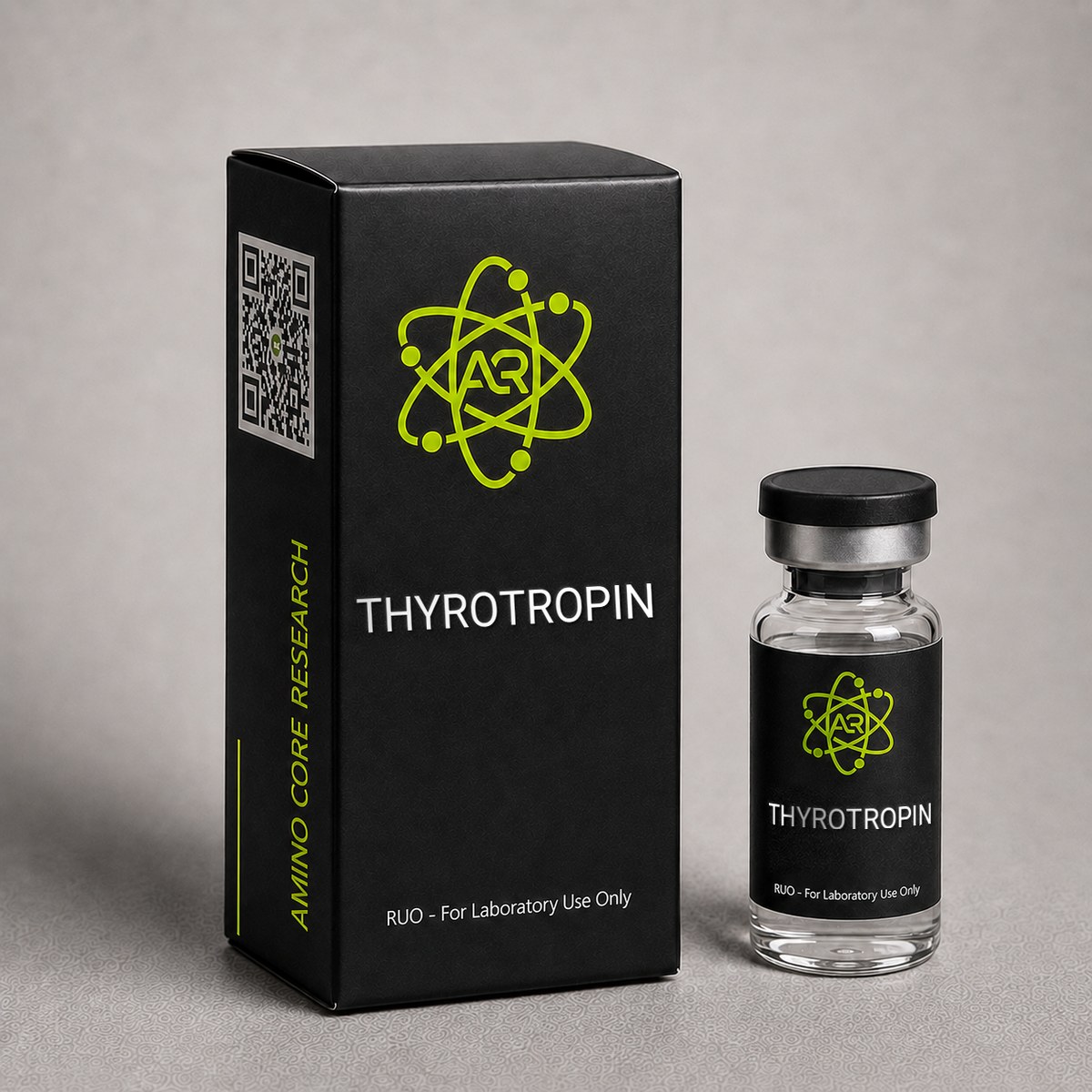
≥99%HPLC Purity
COAIncluded
USAShipped
Thyrotropin (TSH) Peptide
Thyroid-stimulating hormone (TSH) glycoprotein analog for endocrine system research. Key compound in hypothalamic-pituitary-thyroid axis studies and thyroid function research.
Research Reference
This page is a research monograph. Thyrotropin (TSH) is documented here for reference — structure, mechanism and published literature — and is not offered for sale.
Browse compounds available for research purchase · Read the research library
Quick Facts
| SKU | TSH-001 |
|---|---|
| CAS Number | 9002-71-5 |
| Molecular Formula | C1539H2406N418O474S20 |
| Molecular Weight | 28,000 Da |
| Sequence | Alpha subunit (92 aa): APDVQDCPECTLQENPFFSQPGAPILQCMGCCFSRAYPTPLRSKKTMLVQKNVTSESTCCVAKSYNRVTVMGGFKVENHTACHCSTCYYHKS; Beta subunit (118 aa): FCIPTEYTMHIERRECAYCLTINTTICAGYCMTRDINGKLFLPKYALSQDVCTYRDFIYRTVEIPGCPLHVAPYFSYPVALSCKCGKCNTDYSDCIHEAIKTNYCTKPQKSYLVGFSV |
| Purity | ≥99% |
| Physical Form | Lyophilized Powder |
| Storage | Store at -20°C |
What is Thyrotropin (TSH)?
Thyroid-stimulating hormone is a glycoprotein produced by thyrotrope cells in the anterior pituitary. It binds the TSH receptor (TSHR) on thyroid follicular cells, activating cAMP/PKA signaling to stimulate thyroglobulin synthesis, iodide uptake (NIS), and T3/T4 hormone release. Essential compound for hypothalamic-pituitary-thyroid axis research.
Mechanism of Action
Thyrotropin (TSH) is a heterodimeric glycoprotein hormone composed of an alpha subunit shared with LH, FSH, and hCG, and a unique beta subunit that confers receptor specificity. Its biological activity is mediated through binding to the thyroid-stimulating hormone receptor (TSHR), a G-protein-coupled receptor (GPCR) of the rhodopsin-like family expressed predominantly on the basolateral membrane of thyroid follicular cells.
TSH Receptor Activation
Upon TSH binding to the large extracellular leucine-rich repeat domain of TSHR, conformational changes propagate through the transmembrane hinge region, activating coupled heterotrimeric G-proteins. TSHR is unique among glycoprotein hormone receptors in its promiscuous G-protein coupling, activating both Gs-alpha and Gq/11 pathways depending on receptor occupancy and cellular context.
cAMP/PKA Pathway
The dominant signaling cascade involves Gs-alpha activation of adenylyl cyclase, increasing intracellular cyclic AMP (cAMP). Elevated cAMP activates protein kinase A (PKA), which phosphorylates transcription factors including CREB. This drives expression of thyroid-specific genes encoding thyroglobulin (Tg), thyroid peroxidase (TPO), the sodium-iodide symporter (NIS), and dual oxidase (DUOX). The net result is increased iodide uptake, organification, and synthesis of thyroxine (T4) and triiodothyronine (T3).
Phospholipase C/PKC Pathway
At higher TSH concentrations, Gq/11 coupling activates phospholipase C-beta, generating inositol trisphosphate (IP3) and diacylglycerol (DAG). This mobilizes intracellular calcium and activates protein kinase C (PKC), modulating H2O2 generation via DUOX2 — essential for iodide organification and hormone coupling reactions catalyzed by TPO.
Growth and Differentiation Effects
Chronic TSH stimulation activates PI3K/AKT and MAPK/ERK pathways downstream of cAMP-PKA crosstalk, promoting thyrocyte proliferation, hypertrophy, and vascularization of the thyroid gland. Sustained elevated TSH in research models drives goiter formation, while TSHR loss-of-function results in thyroid hypoplasia.
Negative Feedback Regulation
Circulating T3 and T4 exert negative feedback at both the hypothalamus (suppressing TRH) and anterior pituitary thyrotrophs (suppressing TSH transcription and secretion). T3 binds thyroid hormone receptor beta-2 (TR-beta-2) in thyrotrophs, repressing the TSH-beta and alpha-subunit genes. This closed-loop architecture maintains euthyroid homeostasis.
Extrathyroidal TSHR Signaling
TSHR expression has been documented in adipocytes, osteoblasts, fibroblasts, lymphocytes, and orbital tissue. In retro-orbital fibroblasts, TSHR cross-talks with IGF-1R signaling — a mechanism studied extensively in Graves' orbitopathy research models. In bone, TSHR signaling appears to modulate osteoclast differentiation, suggesting a direct role beyond thyroid hormone-mediated effects.
Research & Clinical Studies
Recombinant Human TSH in Thyroid Cancer Research Models
Recombinant human thyrotropin (rhTSH) has been extensively studied as a stimulation agent in differentiated thyroid cancer (DTC) research, enabling thyroglobulin testing and radioiodine uptake without thyroid hormone withdrawal in preclinical and clinical models.
Pivotal Stimulation Studies
Foundational work by Ladenson and colleagues evaluated rhTSH administration in subjects with previously treated DTC. The investigational protocol used two intramuscular doses of 0.9 mg rhTSH on consecutive days, with thyroglobulin measurement and radioiodine scanning 72 hours after the second dose.
- rhTSH stimulation elevated serum TSH to a mean peak of 132 mU/L within 24 hours of the second dose
- Detection of residual or recurrent thyroid tissue was concordant with thyroid hormone withdrawal in ~89% of cases when combined with thyroglobulin measurement
- Quality-of-life metrics remained stable during rhTSH stimulation versus significant deterioration during hormone withdrawal
- No anti-TSH antibody formation was detected following repeat administration cycles
Mechanistic Validation
The studies confirmed that exogenous TSH can drive functional expression of NIS in differentiated thyroid carcinoma tissue, enabling radioiodine concentration sufficient for both diagnostic imaging and ablative protocols. This provided proof-of-concept that TSHR signaling remains intact in well-differentiated tumors and can be pharmacologically exploited.
Extended Research Applications
Subsequent investigations demonstrated rhTSH-stimulated thyroglobulin measurement achieves diagnostic sensitivity of approximately 92-96% for detecting persistent or recurrent disease when combined with neck ultrasonography. The research established serum thyroglobulin thresholds of >2 ng/mL post-stimulation as indicative of clinically relevant residual tissue.
Comparative pharmacokinetic studies show rhTSH has an elimination half-life of approximately 22-35 hours, with peak serum concentrations occurring 3-24 hours after intramuscular injection. Renal clearance is the predominant elimination route, and dose adjustments have been studied in models of impaired renal function.
[1] Ladenson PW, Braverman LE, Mazzaferri EL, et al. Comparison of administration of recombinant human thyrotropin with withdrawal of thyroid hormone for radioactive iodine scanning in patients with thyroid carcinoma. N Engl J Med. 1997;337(13):888-896. PubMed ↗
[2] Haugen BR, Pacini F, Reiners C, et al. A comparison of recombinant human thyrotropin and thyroid hormone withdrawal for the detection of thyroid remnant or cancer. J Clin Endocrinol Metab. 1999;84(11):3877-3885. PubMed ↗
Recombinant Human TSH and Radioiodine Uptake in Thyroid Cancer Research
One of the most extensively studied applications of recombinant human thyrotropin (rhTSH, thyrotropin alfa) in clinical and translational research is its use to enhance radioiodine (I-131) uptake and thyroglobulin secretion in differentiated thyroid cancer (DTC) follow-up models. Prior to rhTSH availability, researchers and clinicians relied exclusively on thyroid hormone withdrawal (THW) to elevate endogenous TSH, a method that introduced significant physiological stress and confounding variables into study designs.
A landmark multicenter investigation published by Ladenson and colleagues compared rhTSH-stimulated radioiodine scanning to traditional THW protocols in 127 patients with DTC. The study reported that rhTSH administration (0.9 mg intramuscularly on two consecutive days) produced serum TSH concentrations comparable to those achieved by hypothyroidism induction, with peak TSH levels exceeding 130 mIU/L within 24 hours of the second dose. Critically, the agreement between rhTSH and THW scans for detecting residual or metastatic thyroid tissue was 89%, and stimulated thyroglobulin measurements showed strong concordance between the two methods.
Key findings reported across follow-up studies include:
- Sensitivity of rhTSH-stimulated thyroglobulin for detecting persistent or recurrent disease: ~85-95% when combined with neck ultrasound.
- Radioiodine uptake in residual thyroid tissue: equivalent or superior to THW in approximately 80% of paired comparisons.
- Patient-reported quality of life scores were significantly higher with rhTSH preparation versus THW (p<0.001), eliminating the confound of hypothyroid symptomatology in metabolic substudies.
- Pharmacokinetics: elimination half-life of approximately 25 hours, allowing stable TSH receptor engagement during the scanning window.
Subsequent research has validated rhTSH as a standardized tool for probing TSH receptor function in vivo without disturbing systemic thyroid hormone homeostasis. This property has made rhTSH particularly valuable in studies examining extra-thyroidal TSH receptor expression in bone, adipose tissue, and immune cells, where investigators require a controlled TSH stimulus uncoupled from changes in T3/T4 levels. Comparative research suggests that rhTSH provides a more reproducible pharmacological stimulus than endogenous TSH elevation, with lower inter-subject variability in peak hormone concentrations.
In preclinical models, recombinant TSH has been used to characterize sodium-iodide symporter (NIS) upregulation, thyroglobulin gene transcription, and follicular cell proliferation kinetics. These investigations have informed the broader understanding of TSH receptor pharmacology and continue to shape research into TSH receptor-targeted theranostics and TSH receptor-mediated disease models such as Graves' disease and toxic nodular goiter.
[1] Ladenson PW, Braverman LE, Mazzaferri EL, et al. Comparison of administration of recombinant human thyrotropin with withdrawal of thyroid hormone for radioactive iodine scanning in patients with thyroid carcinoma. N Engl J Med. 1997;337(13):888-896. PubMed ↗
[2] Haugen BR, Pacini F, Reiners C, et al. A comparison of recombinant human thyrotropin and thyroid hormone withdrawal for the detection of thyroid remnant or cancer. J Clin Endocrinol Metab. 1999;84(11):3877-3885. PubMed ↗
TSH Receptor Signaling and Extrathyroidal Effects in Bone Research Models
Beyond its classical role in regulating thyroid follicular cells, thyrotropin has become an important research tool for investigating TSH receptor (TSHR) signaling in extrathyroidal tissues. A pivotal study by Abe and colleagues published in Cell demonstrated that TSHR is expressed on osteoblasts and osteoclasts, and that TSH acts as a direct negative regulator of skeletal remodeling, independent of thyroid hormone levels. This finding reframed the interpretation of long-standing clinical observations linking suppressed TSH to bone loss.
In the foundational mouse model, TSHR-null (TSHR-/-) animals exhibited profound osteoporosis with both increased bone resorption and decreased bone formation, despite thyroid hormone replacement to euthyroid levels. Heterozygous TSHR+/- mice, which had only modestly elevated TSH compensation, also displayed bone loss, demonstrating a gene-dose-dependent effect of TSHR signaling on skeletal homeostasis. Key mechanistic findings included:
- TNF-alpha production by osteoclast precursors increased ~3-fold in TSHR-deficient cells, identifying a pro-resorptive cytokine pathway suppressed by physiological TSH signaling.
- Osteoblast differentiation markers (Runx2, osterix, type I collagen) were reduced by 40-60% in the absence of TSHR signaling.
- Low-dose recombinant TSH administration in ovariectomized rat models produced a ~25% increase in trabecular bone volume over 8 weeks without altering circulating thyroid hormone concentrations.
Subsequent investigations have used recombinant thyrotropin as a probe to dissect TSHR-mediated cAMP and Gq-coupled signaling in primary osteoblast cultures, bone marrow stromal cells, and adipocyte differentiation models. Research has identified that TSHR activation modulates Wnt/beta-catenin signaling and influences mesenchymal stem cell fate decisions between osteoblastic and adipocytic lineages.
Additional extrathyroidal TSHR research has examined:
- Adipose tissue: TSHR expression on preadipocytes and mature adipocytes, with TSH stimulating lipolysis and IL-6 release in cultured human adipocytes.
- Cardiac tissue: low-level TSHR expression with reported effects on cardiomyocyte contractility in isolated preparations.
- Immune cells: TSHR expression on dendritic cells and lymphocytes, with implications for autoimmune thyroid disease research (Graves' disease, Hashimoto's thyroiditis).
These extrathyroidal investigations have established recombinant TSH as an indispensable research reagent for studying pleiotropic GPCR signaling. Because endogenous TSH is normally tightly coupled to systemic thyroid hormone status, exogenous rhTSH administration provides researchers with a unique pharmacological tool to isolate TSHR-specific effects from secondary thyroid hormone-mediated changes.
Chemical & Physical Properties
Thyrotropin is a heterodimeric glycoprotein hormone with extensive post-translational modifications including N-linked glycosylation critical for receptor binding, bioactivity, and circulatory half-life. The following table summarizes verified physicochemical properties of recombinant human TSH used in research applications.
| Full Name | Thyroid-Stimulating Hormone / Thyrotropin |
|---|---|
| Synonyms | TSH, hTSH, Thyrotropin Alfa (recombinant), Thyrogen |
| Molecular Formula | C₁₅₃₉H₂₄₀₆N₄₁₈O₄₇₄S₂₀ (protein backbone, excluding glycans) |
| Molecular Weight | ~28,000 Da (glycosylated heterodimer; ~22 kDa peptide backbone) |
| CAS Number | 9002-71-5 |
| UniProt IDs | P01215 (alpha subunit), P01222 (beta subunit) |
| Subunit Composition | Non-covalent heterodimer: alpha subunit (92 aa) + beta subunit (118 aa) |
| Amino Acid Count | 210 total residues (alpha 92 + beta 118) |
| Disulfide Bonds | 5 in alpha subunit, 6 in beta subunit (cystine knot motif) |
| Glycosylation | Two N-glycosylation sites on alpha subunit (Asn52, Asn78); one on beta subunit (Asn23) |
| Receptor Target | Thyroid-Stimulating Hormone Receptor (TSHR / LGR3) |
| Origin / Developer | Endogenous: anterior pituitary thyrotrophs. Recombinant: CHO cell expression (Genzyme, 1998) |
| Hormone Family | Glycoprotein hormone family (with LH, FSH, hCG) |
| Physical Form | Lyophilized powder |
| Solubility | Soluble in sterile water for injection, bacteriostatic water, or PBS |
| Purity | ≥99% by HPLC |
| Isoelectric Point | ~7.5 (variable due to sialic acid content) |
The shared alpha subunit is identical to that of luteinizing hormone (LH), follicle-stimulating hormone (FSH), and human chorionic gonadotropin (hCG). Biological specificity is conferred entirely by the unique beta subunit, which determines receptor binding selectivity. The cystine knot architecture stabilizes the dimer through three interlocking disulfide bonds in each subunit, making TSH highly resistant to denaturation under physiological conditions.
Sialic acid content on the carbohydrate moieties governs circulatory half-life: more highly sialylated TSH isoforms exhibit longer plasma persistence, while less sialylated forms have shorter half-life but higher in vitro receptor potency. This glycoform heterogeneity is a key consideration in pharmacokinetic research models.
Handling & Reconstitution Guidelines
Thyrotropin (TSH) is a heterodimeric glycoprotein with two non-covalently associated subunits (alpha and beta) stabilized by multiple intrachain disulfide bonds and N-linked glycosylation. Proper handling is critical to preserve heterodimer integrity, glycan structure, and TSH receptor binding activity. The lyophilized form is supplied as a sterile powder, typically with mannitol or sucrose as a lyoprotectant.
Recommended Reconstitution Protocol:
- Equilibrate the vial to room temperature (15-25 degrees C) for 15-20 minutes before opening. Do not warm above 25 degrees C, as elevated temperatures can promote subunit dissociation and aggregation.
- Select an appropriate diluent. Sterile bacteriostatic water for injection (0.9% benzyl alcohol) or sterile sodium chloride 0.9% is commonly used. For sensitive cell culture work, sterile sodium chloride is preferred to avoid benzyl alcohol cytotoxicity.
- Calculate working concentration. A typical reconstitution is 0.9 mg of lyophilized rhTSH + 1.2 mL diluent, yielding a working concentration of approximately 0.9 mg/1.2 mL (0.75 mg/mL). Other concentrations (e.g., 100 micrograms/mL for cell culture stimulation) can be prepared as needed.
- Add diluent slowly down the inner wall of the vial. Do not direct the stream onto the lyophilized cake.
- Dissolve by gentle swirling for 30-60 seconds. Do NOT vortex or shake vigorously — mechanical agitation can shear the glycoprotein heterodimer and generate inactive aggregates that retain immunoreactivity but lack receptor binding activity.
- Inspect visually. The reconstituted solution should be clear and colorless. Discard if cloudy, particulate, or discolored.
- Aliquot promptly into single-use sterile tubes (low-binding polypropylene recommended) to minimize freeze-thaw cycles.
Compound-Specific Handling Notes:
- Glycosylation sensitivity: Avoid exposure to extremes of pH (<5 or >9), which can promote deglycosylation and loss of bioactivity.
- Adsorption losses: At low concentrations (<10 micrograms/mL), TSH can adsorb to glass and standard polypropylene surfaces. Carrier protein (0.1% BSA) is recommended for dilute working solutions used in receptor binding assays.
- Light exposure: While not strongly photolabile, prolonged exposure to direct light should be avoided to minimize oxidative damage to methionine and tryptophan residues.
- Aseptic technique: Always use sterile filtered diluents and work in a laminar flow hood for cell culture applications.
This product is intended for in vitro laboratory research use only and is not for human or veterinary administration.
Frequently Asked Questions
What does TSH stimulate in the thyroid?
TSH activates thyroid follicular cells to: (1) increase iodide uptake via NIS transporter, (2) synthesize thyroglobulin, (3) produce and release T4 (thyroxine) and T3 (triiodothyronine), (4) promote thyroid cell growth.
What is the molecular weight and structure of Thyrotropin (TSH)?
Thyrotropin has an approximate molecular weight of 28,000 Da (28 kDa) as a fully glycosylated heterodimer, with CAS number 9002-71-5. It consists of two non-covalently associated subunits: an alpha subunit of 92 amino acids (shared with LH, FSH, and hCG) and a unique beta subunit of 118 amino acids that confers receptor specificity. The molecule contains 11 disulfide bonds arranged in cystine knot motifs and three N-linked glycosylation sites. Glycosylation contributes approximately 6 kDa to the total mass and is essential for circulatory half-life and receptor binding.
How does Thyrotropin (TSH) compare to TRH in endocrine research?
Thyrotropin (TSH) and thyrotropin-releasing hormone (TRH) act at different levels of the hypothalamic-pituitary-thyroid axis. TRH is a small tripeptide (pGlu-His-Pro-NH2, MW ~362 Da) released from the hypothalamus that stimulates pituitary thyrotrophs to synthesize and secrete TSH. TSH, a 28 kDa glycoprotein, then acts directly on thyroid follicular cells via the TSH receptor (TSHR) to drive T3/T4 production. In research models, TRH is used to probe pituitary thyrotroph function, while TSH is used to directly stimulate thyroid tissue, including in radioiodine uptake studies where bypassing the hypothalamic-pituitary stage is required.
How should Thyrotropin (TSH) be stored for research use?
Lyophilized Thyrotropin (TSH) should be stored at -20°C for long-term stability, where it remains stable for 24+ months. Short-term storage at 2-8°C is acceptable for several weeks. Transit at room temperature for up to one week does not compromise integrity due to the stabilizing disulfide architecture. Once reconstituted in sterile diluent, TSH should be stored at 2-8°C and used within 24 hours, or aliquoted and frozen at -20°C for up to 4 weeks. Avoid repeated freeze-thaw cycles, which can disrupt the heterodimer association and reduce bioactivity. Protect from light and avoid vigorous agitation.
What research applications use Thyrotropin (TSH)?
Thyrotropin (TSH) is used in research models to study thyroid follicular cell function, TSH receptor (TSHR) signaling, iodide transport via the sodium-iodide symporter (NIS), and thyroid hormone biosynthesis. It serves as a stimulation agent in differentiated thyroid cancer research to enhance radioiodine uptake and thyroglobulin expression without thyroid hormone withdrawal. TSH is also employed in extrathyroidal TSHR research, including studies of orbital fibroblast cross-talk with IGF-1R (relevant to Graves' orbitopathy models), adipocyte biology, and bone remodeling investigations. Pharmacokinetic studies use TSH to model glycoprotein hormone clearance and bioavailability across glycoform variants.
What is the half-life of recombinant Thyrotropin (TSH) in pharmacokinetic studies?
Recombinant human thyrotropin (rhTSH) demonstrates an elimination half-life of approximately 22-35 hours following intramuscular administration in pharmacokinetic studies. Peak serum TSH concentrations are typically reached within 3-24 hours after a 0.9 mg dose, with peak levels often exceeding 130 mIU/L. The relatively long half-life reflects the heavily glycosylated nature of the heterodimeric glycoprotein, which slows renal clearance compared to non-glycosylated peptide hormones. This sustained pharmacological exposure allows researchers to maintain TSH receptor engagement throughout a multi-day stimulation protocol, making rhTSH particularly useful for radioiodine uptake studies and TSH receptor signaling investigations.
Does Thyrotropin (TSH) affect bone tissue independent of thyroid hormone?
Research published in Cell by Abe and colleagues (2003) demonstrated that TSH acts directly on bone cells through TSH receptor (TSHR) expression on osteoblasts and osteoclasts, independent of thyroid hormone effects. TSHR-knockout mice exhibited profound osteoporosis even when maintained at euthyroid thyroid hormone levels, with increased osteoclast TNF-alpha production and decreased osteoblast differentiation markers. Subsequent studies showed that recombinant TSH administration in ovariectomized rat models increased trabecular bone volume by approximately 25% over 8 weeks without altering circulating T3 or T4. These findings establish TSH as a direct skeletal regulator and have made rhTSH a valuable research tool for studying extrathyroidal GPCR signaling.
What is the difference between native pituitary TSH and recombinant human TSH (rhTSH)?
Native pituitary TSH and recombinant human TSH (rhTSH, thyrotropin alfa) share the same amino acid sequence for both alpha and beta subunits but differ in glycosylation patterns. Native TSH is glycosylated by pituitary thyrotroph cells, producing a heterogeneous mix of glycoforms with varying sialic acid content. Recombinant TSH is typically produced in Chinese hamster ovary (CHO) cells, yielding a more uniform glycoform with slightly different sialylation. These glycan differences influence circulating half-life and receptor binding kinetics, with rhTSH generally showing more reproducible pharmacokinetics in research applications. Both forms activate TSH receptors with comparable potency in vitro.
Why must Thyrotropin (TSH) not be vortexed during reconstitution?
Thyrotropin is a non-covalently associated heterodimer of alpha and beta subunits stabilized by hydrophobic and ionic interactions plus extensive glycosylation. Vigorous mechanical agitation such as vortexing introduces shear forces and air-liquid interface stress that can dissociate the subunits and promote irreversible aggregation. Aggregated TSH may retain immunoreactivity in ELISA-based assays but loses biological activity at the TSH receptor, leading to falsely reduced bioactivity in research preparations. The correct technique is gentle swirling for 30-60 seconds at room temperature, allowing the lyophilized cake to dissolve without mechanical disruption of the glycoprotein structure.
For laboratory and research use only. This product is not for human or veterinary use. It is not a drug, supplement or food.
This product is not FDA approved for any indication, and is not intended to diagnose, treat, cure, or prevent any disease. All product information is derived from published preclinical research and does not constitute medical advice or claims.