≥99%HPLC Purity
COAIncluded
USAShipped
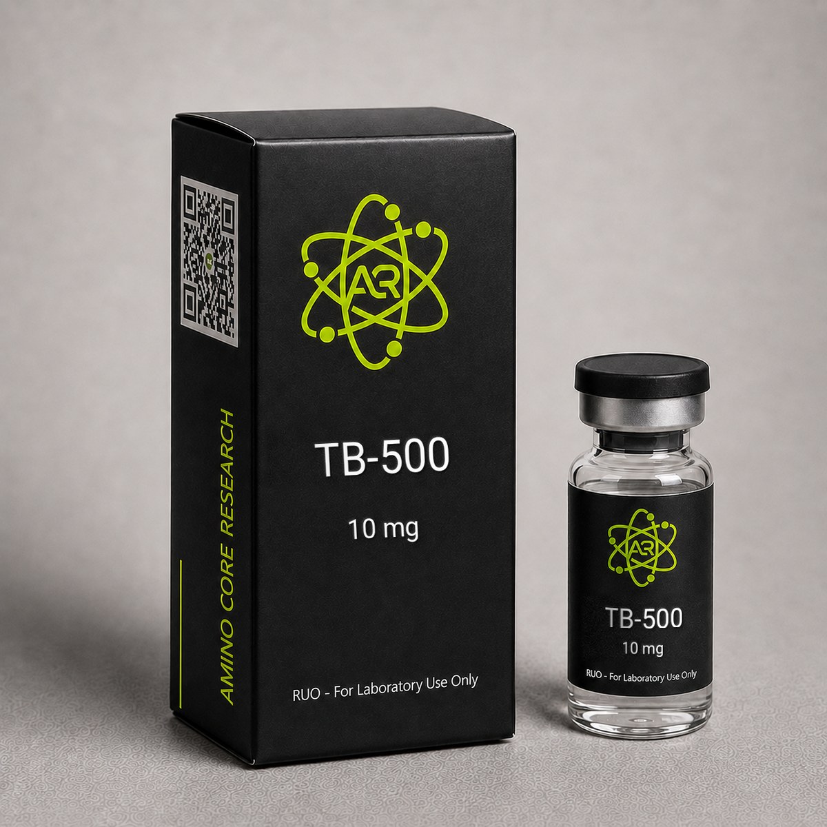
TB-500 Peptide
Thymosin Beta-4 fragment peptide. A 43-amino acid peptide originally isolated from thymus tissue, studied extensively in cell biology research.
$99.00
In stock — ships next business day
Quick Facts
| SKU | ACR-TB500 |
|---|---|
| CAS Number | 77591-33-4 |
| Molecular Formula | C212H350N56O78S |
| Molecular Weight | 4963.44 g/mol |
| Sequence | 43-amino acid sequence |
| Purity | ≥99% |
| Physical Form | Lyophilized Powder |
| Storage | Store at -20°C |
What is TB-500?
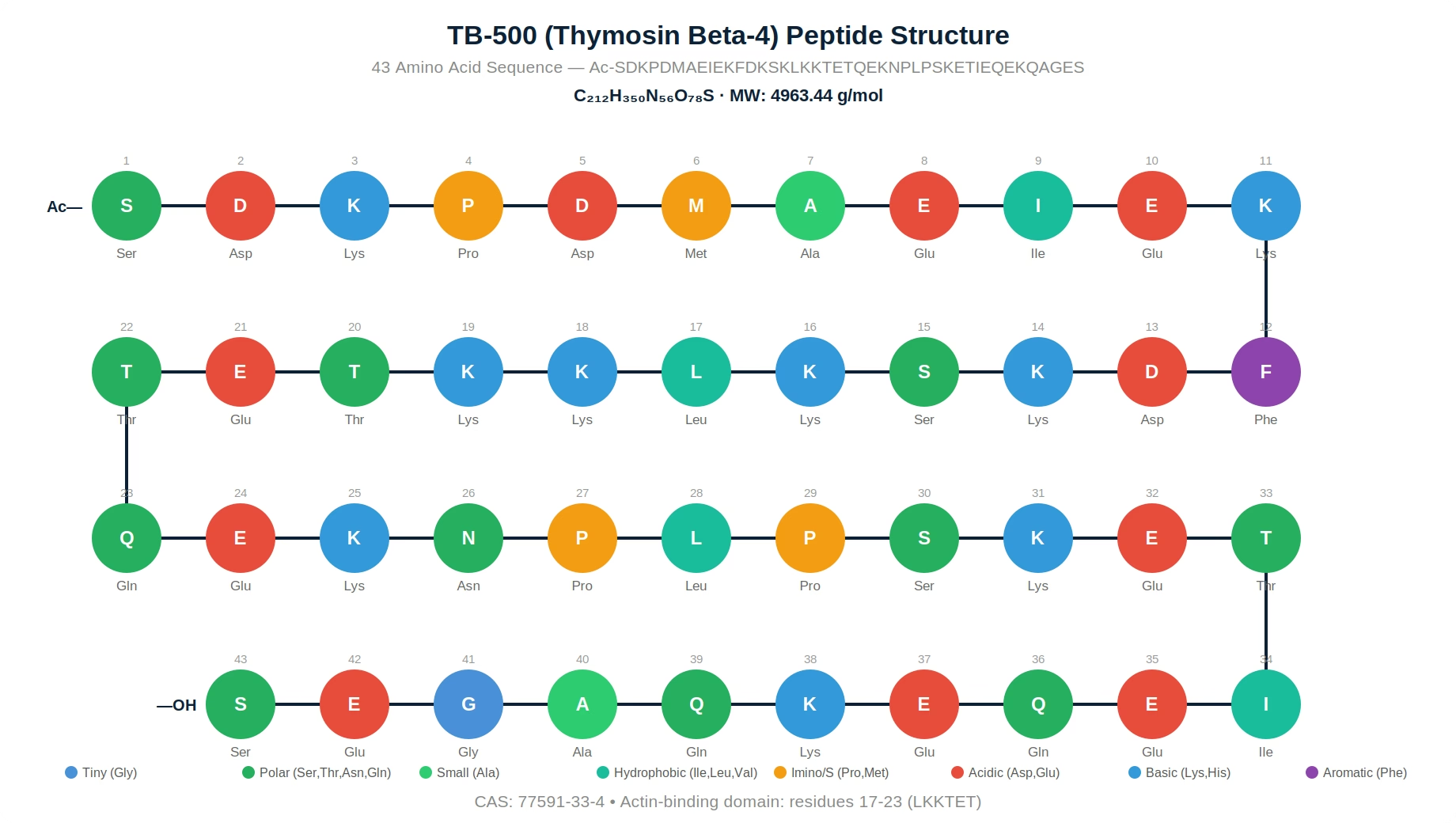
TB-500 is a synthetic version of Thymosin Beta-4 (Tβ4), a naturally occurring 43-amino acid peptide present in virtually all human and animal cells, with the notable exception of red blood cells. Thymosin Beta-4 was first isolated from calf thymus tissue by Allan Goldstein and colleagues in the 1960s as part of a broader effort to characterize thymic peptides involved in immune function. The peptide has the amino acid sequence Ac-SDKPDMAEIEKFDKSKLKKTETQEKNPLPSKETIEQEKQAGES, with a molecular weight of 4963.44 g/mol and CAS number 77591-33-4.
Thymosin Beta-4 is the most abundant member of the beta-thymosin family and constitutes up to 70-80% of the total actin-sequestering activity in most cell types. Intracellular concentrations can reach 100-500 μM in highly motile cells such as platelets, macrophages, and neutrophils. Its primary biological role is regulation of the actin cytoskeleton — the dynamic structural framework that enables cells to move, divide, and change shape.
Beyond its cytoskeletal functions, published research over the past two decades has revealed that Tβ4 has significant extracellular signaling roles. When released from cells (particularly platelets during clotting), extracellular Tβ4 appears to activate signaling pathways involved in tissue repair, angiogenesis, and inflammation modulation. This dual intracellular/extracellular functionality makes Tβ4 one of the most versatile peptides characterized in the published literature.
TB-500 is classified as a research peptide and is available exclusively for laboratory and scientific investigation. All findings described herein are derived from published preclinical and clinical studies and do not constitute medical claims.
Mechanism of Action
Published research has identified several key molecular pathways through which Thymosin Beta-4 exerts its biological effects. The peptide appears to operate through both intracellular mechanisms (actin regulation) and extracellular signaling pathways (receptor-mediated).
Actin Sequestration and Cytoskeletal Dynamics
The primary intracellular function of Tβ4 is binding monomeric G-actin (globular actin) in a 1:1 complex, preventing its spontaneous polymerization into F-actin (filamentous actin) filaments. The actin-binding domain spans residues 17-23 of the peptide, with the key motif LKKTET making direct contact with the actin monomer. This sequestration maintains a large reservoir of polymerization-ready actin monomers that can be rapidly mobilized when cells receive migration signals. The dynamic equilibrium between Tβ4-bound G-actin and profilin-bound G-actin (which promotes polymerization) is a critical determinant of cell motility.
Integrin-Linked Kinase (ILK) / Akt Pathway
Bock-Marquette et al. (2004) demonstrated that Tβ4 activates integrin-linked kinase (ILK), a serine/threonine kinase that connects extracellular matrix signals to intracellular cascades. ILK activation leads to phosphorylation of Akt (protein kinase B) at Ser473, activating a major cell survival and migration pathway. Downstream Akt targets include Bad (pro-apoptotic protein inactivation), GSK-3β (glycogen synthase kinase inhibition), and mTOR (protein synthesis promotion).
Matrix Metalloproteinase (MMP) Regulation
Published studies have documented that Tβ4 increases expression of several matrix metalloproteinases, particularly MMP-2 and MMP-9. These zinc-dependent endopeptidases degrade extracellular matrix components, creating pathways for cell migration during tissue remodeling. MMP regulation appears to be mediated through NF-κB and AP-1 transcription factor activation.
Anti-inflammatory Signaling
Research indicates that Tβ4 may modulate inflammatory responses through several mechanisms: suppression of NF-κB nuclear translocation, reduction of pro-inflammatory cytokine expression (TNF-α, IL-1β, IL-6), and promotion of anti-inflammatory mediators. The sulfoxide form of Tβ4 (Tβ4-SO) has been specifically studied for anti-inflammatory signaling through inhibition of NF-κB.
Angiogenesis Promotion
Tβ4 promotes endothelial cell differentiation and new blood vessel formation. Published research has shown increased expression of vascular endothelial growth factor (VEGF) and its receptors in Tβ4-treated tissues, along with enhanced endothelial cell tubule formation in Matrigel assays. The angiogenic effect appears to involve both Akt signaling and direct effects on endothelial progenitor cell recruitment.
Research & Clinical Studies
TB-500 and Wound Healing Research
Malinda et al. (1999) published one of the foundational studies on Thymosin Beta-4 in wound healing using dermal wound models. The study utilized full-thickness excisional wound models in rats to assess the effects of topical and systemic Tβ4 administration.
The researchers reported several key findings: Tβ4-treated wounds showed accelerated closure rates compared to saline controls, with statistically significant differences observed at days 4, 7, and 11 post-injury. Histological analysis revealed increased collagen deposition (primarily type I and III collagen), enhanced angiogenesis with greater capillary density in the wound bed, and accelerated re-epithelialization with organized stratification of the neoepidermis.
In parallel in-vitro experiments, the study demonstrated that Tβ4 promoted keratinocyte migration in scratch wound assays at concentrations as low as 1 μg/mL. Endothelial cell tubule formation on Matrigel matrices was enhanced 2-3 fold compared to controls. Importantly, Tβ4 did not stimulate cell proliferation in these models, suggesting its primary wound healing mechanism involves migration rather than mitogenic signaling.
A subsequent study by Philp et al. (2004) extended these findings using a corneal wound model. The researchers demonstrated that Tβ4 promoted corneal epithelial cell migration and wound closure, with apparent anti-inflammatory effects including reduced polymorphonuclear leukocyte infiltration. The corneal model provided a well-defined system for quantifying epithelial migration rates.
[1] Malinda KM, et al. Thymosin beta4 accelerates wound healing. J Invest Dermatol. 1999;113(3):364-8.
[2] Philp D, et al. Thymosin beta4 promotes angiogenesis, wound healing, and hair follicle development. Mech Ageing Dev. 2004;125(2):113-5. PubMed ↗
TB-500 and Cardiac Tissue Research
Bock-Marquette et al. (2004) published a landmark study in Nature demonstrating that Thymosin Beta-4 plays a critical role in cardiac tissue biology. This study fundamentally expanded understanding of Tβ4 beyond its cytoskeletal functions into tissue-protective signaling.
The researchers first established that Tβ4 is highly expressed in embryonic cardiomyocytes during heart development. Using in-vitro cardiomyocyte cultures subjected to hypoxic stress, they demonstrated that Tβ4 treatment promoted cell survival by activating integrin-linked kinase (ILK). ILK activation led to Akt phosphorylation at Ser473, which in turn inactivated the pro-apoptotic protein Bad and activated cell survival pathways.
In the in-vivo component, murine models with experimentally induced myocardial injury were treated with systemic Tβ4. The treated group showed significant preservation of cardiac function as measured by echocardiography. Specifically, ejection fraction and fractional shortening were improved in the Tβ4 group compared to controls. Histological analysis revealed reduced infarct size and decreased TUNEL-positive (apoptotic) cardiomyocytes.
Smart et al. (2007) followed up with a critical finding: Tβ4 could reactivate quiescent epicardial progenitor cells in adult hearts. These progenitor cells, marked by the transcription factor Wt1, migrated into the myocardium and appeared to differentiate into new cardiomyocytes and vascular cells. This suggested that Tβ4 might activate endogenous cardiac regeneration rather than merely protecting existing cells.
[3] Bock-Marquette I, et al. Thymosin beta4 activates integrin-linked kinase and promotes cardiac cell migration, survival and cardiac repair. Nature. 2004;432(7016):466-72. PubMed ↗
[4] Smart N, et al. Thymosin beta4 induces adult epicardial progenitor mobilization and neovascularization. Nature. 2007;445(7124):177-82. PubMed ↗
TB-500 and Corneal Repair Research
Sosne et al. (2002, 2005) conducted a series of studies examining Thymosin Beta-4 in corneal tissue repair models. The cornea provides an ideal tissue for studying wound healing peptides due to its accessibility, transparency (allowing real-time monitoring), and well-characterized healing phases.
In the initial study, Sosne et al. (2002) demonstrated that Tβ4 promoted corneal epithelial cell migration in a dose-dependent manner using both scratch wound and transwell migration assays. The peptide did not increase cell proliferation, confirming a pro-migratory rather than mitogenic mechanism. Concentrations between 0.1-10 μg/mL showed optimal activity.
The 2005 follow-up study utilized an alkali burn model, which produces severe corneal damage including stromal degradation and persistent inflammation. Tβ4-treated eyes showed accelerated epithelial recovery, reduced corneal haze (indicating less fibrotic scarring), and decreased inflammatory cell infiltration. Importantly, the researchers identified that Tβ4 decreased MMP-2 and MMP-9 activity in the damaged cornea — apparently contradicting findings in other tissues where Tβ4 increases MMP expression. This suggested tissue-specific regulation, where Tβ4 promotes controlled MMP activity for migration but prevents excessive proteolytic destruction in severely damaged tissue.
These corneal studies were significant because they demonstrated that Tβ4 effects are context-dependent, promoting orderly tissue repair rather than simply activating a single pathway universally.
[5] Sosne G, et al. Thymosin beta 4 promotes corneal wound healing and decreases inflammation in vivo following alkali injury. Exp Eye Res. 2002;74(2):293-9. PubMed ↗
[6] Sosne G, et al. Thymosin beta4 promotes corneal wound healing and modulates inflammatory mediators in vivo. Exp Eye Res. 2005;81(5):583-8. PubMed ↗
TB-500 and Inflammatory Response Research
The anti-inflammatory properties of Thymosin Beta-4 have been investigated across multiple experimental systems. Young et al. (1999) first demonstrated that Tβ4 could reduce the expression of pro-inflammatory chemokines in stimulated cell cultures, suggesting direct modulation of inflammatory gene transcription.
Subsequent research by Sosne et al. (2007) investigated the anti-inflammatory mechanism in greater detail. Using TNF-α-stimulated human corneal epithelial cells, the researchers showed that Tβ4 inhibited NF-κB activation by preventing IκB-α degradation and subsequent p65 nuclear translocation. This NF-κB inhibition reduced expression of downstream inflammatory mediators including IL-1β, IL-6, IL-8, and TNF-α itself, creating a negative feedback loop.
The sulfoxide derivative of Tβ4 (Tβ4-SO), formed by oxidation of the Met6 residue, has been specifically studied for anti-inflammatory signaling. Published research suggests Tβ4-SO may have enhanced anti-inflammatory activity compared to the reduced form, particularly in neutrophil-mediated inflammation models. This is physiologically relevant because the inflammatory environment is highly oxidative, potentially converting Tβ4 to its sulfoxide form at sites of tissue damage.
[7] Young JD, et al. Thymosin beta 4 sulfoxide is an anti-inflammatory agent generated by monocytes in the presence of glucocorticoids. Nat Med. 1999;5(12):1424-7. PubMed ↗
[8] Sosne G, et al. Thymosin beta 4 modulation of corneal matrix metalloproteinase levels. Invest Ophthalmol Vis Sci. 2005;46:E-2490.
TB-500 and Angiogenesis Research
Grant et al. (1999) published an important study characterizing Thymosin Beta-4 as a potent angiogenic factor. Using human umbilical vein endothelial cells (HUVECs) on Matrigel matrices, the researchers demonstrated that Tβ4 promoted endothelial cell differentiation into tube-like structures — a standard in-vitro measure of angiogenic potential.
The study reported that Tβ4 at concentrations of 1-100 ng/mL induced a 2-3 fold increase in tubule formation compared to controls, with maximal effect at approximately 10 ng/mL. This activity was comparable to established angiogenic factors such as VEGF and bFGF. The researchers also demonstrated that Tβ4 upregulated VEGF expression in endothelial cells, suggesting both direct and indirect angiogenic mechanisms.
Importantly, the angiogenic effect required the intact actin-binding domain of Tβ4. Truncated peptides lacking residues 17-23 (the LKKTET motif) showed significantly reduced angiogenic activity, linking the actin-regulatory function to the angiogenic response. This suggests that Tβ4 promotes angiogenesis at least in part by facilitating endothelial cell migration through cytoskeletal reorganization.
In-vivo confirmation came from chick chorioallantoic membrane (CAM) assays and murine models, where Tβ4 application increased microvessel density and capillary branching in treated areas compared to controls.
[9] Grant DS, et al. Thymosin beta4 enhances endothelial cell differentiation and angiogenesis. Angiogenesis. 1999;3(2):125-35. PubMed ↗
Chemical & Physical Properties
| Property | Value |
|---|---|
| IUPAC Name | Thymosin Beta-4 |
| Molecular Formula | C212H350N56O78S |
| Molecular Weight | 4963.44 g/mol |
| CAS Number | 77591-33-4 |
| Amino Acid Sequence | Ac-SDKPDMAEIEKFDKSKLKKTETQEKNPLPSKETIEQEKQAGES |
| Amino Acid Count | 43 |
| N-Terminal Modification | Acetylated (Ac-Ser) |
| Isoelectric Point (pI) | 5.1 |
| Physical Form | White to off-white lyophilized powder |
| Solubility | Freely soluble in water (>10 mg/mL) |
| Purity | ≥99% (HPLC verified) |
| pH Stability | Stable at pH 4-8 |
| Actin Binding Domain | Residues 17-23 (LKKTET motif) |
| Storage Temperature | -20C (long-term), 2-8C (short-term) |
| Shelf Life | 24 months when stored properly at -20C |
Handling & Reconstitution Guidelines
Reconstitution Protocol
For laboratory use, TB-500 lyophilized powder should be reconstituted using sterile bacteriostatic water (BAC water) or sterile 0.9% sodium chloride solution. Allow the vial to equilibrate to room temperature (approximately 15-20 minutes) before adding diluent. Inject the diluent slowly along the vial wall to avoid creating foam or denaturing the peptide through mechanical stress. Do not vortex or shake vigorously — gently swirl the vial until the powder is fully dissolved. The resulting solution should be clear and colorless without visible particulates.
Recommended Diluent Volumes
For a 5mg vial: Reconstitute with 1.0-2.0 mL of bacteriostatic water, yielding a concentration of 2.5-5.0 mg/mL. For a 10mg vial: Use 2.0 mL for a 5.0 mg/mL concentration. Exact volumes should be determined based on experimental protocol requirements.
Post-Reconstitution Storage
Once reconstituted, the solution should be stored at 2-8C (standard laboratory refrigerator) and used within 30 days. For extended storage beyond 30 days, aliquot the solution into single-use volumes using sterile technique and freeze at -20C. Each aliquot should contain only the volume needed for a single experimental session to avoid repeated freeze-thaw cycles.
Handling Precautions
Use standard laboratory personal protective equipment (PPE) including nitrile gloves and safety glasses. TB-500 is a large peptide (4.9 kDa) that is susceptible to adsorption onto glass surfaces; use low-binding polypropylene microcentrifuge tubes for aliquoting. Work in a clean environment, ideally a laminar flow hood, to minimize contamination risk. Use sterile needles and syringes for all transfers. Do not expose reconstituted solution to temperatures above 25C for extended periods.
Storage & Stability Information
Lyophilized Form (Unreconstituted)
Store lyophilized TB-500 at -20C in a sealed container protected from light and moisture. Under these conditions, the peptide maintains stability and full biological activity for up to 24 months from the manufacture date. Short-term storage at 2-8C (refrigerated) is acceptable for up to 90 days without significant degradation. Avoid exposure to temperatures above 25C for extended periods, as elevated temperatures can accelerate peptide degradation and aggregation.
Reconstituted Solution
Store reconstituted TB-500 solution at 2-8C (standard refrigeration). Use within 30 days of reconstitution when stored continuously at this temperature. For longer preservation, aliquot into single-use volumes using sterile technique and store at -20C. Frozen aliquots maintain stability for approximately 6 months.
Freeze-Thaw Sensitivity
TB-500 is a relatively large peptide (43 amino acids) and is susceptible to degradation from repeated freeze-thaw cycles. Each freeze-thaw cycle can cause ice crystal formation that mechanically disrupts peptide structure and promotes aggregation. Limit freeze-thaw cycles to a maximum of 3 for any given aliquot. For best results, prepare single-use aliquots before initial freezing.
Stability Notes
TB-500 is stable across a moderate pH range (4-8) but shows optimal stability at physiological pH (7.2-7.4). The methionine residue at position 6 (Met6) is susceptible to oxidation, forming the sulfoxide derivative (Tβ4-SO). While Tβ4-SO retains biological activity, minimize exposure to oxidizing conditions (UV light, peroxides, transition metals). Use inert gas (nitrogen or argon) to displace oxygen from vial headspace for long-term storage of reconstituted solutions.
Frequently Asked Questions
What is TB-500?
TB-500 is a synthetic version of Thymosin Beta-4 (Tβ4), a naturally occurring 43-amino acid peptide found in virtually all human and animal cells. Thymosin Beta-4 was first isolated from calf thymus tissue in the 1960s and functions primarily as the major actin-sequestering molecule in eukaryotic cells, regulating cytoskeletal dynamics essential for cell motility, division, and shape changes. Published research has documented significant extracellular signaling roles beyond its cytoskeletal functions, including tissue repair, angiogenesis, and inflammatory response modulation. TB-500 is available exclusively for laboratory and research purposes.
What is the molecular weight and structure of TB-500?
TB-500 has a molecular formula of C212H350N56O78S and a molecular weight of 4963.44 g/mol. The full amino acid sequence is Ac-SDKPDMAEIEKFDKSKLKKTETQEKNPLPSKETIEQEKQAGES (43 residues, N-terminally acetylated). The actin-binding domain is located at residues 17-23 (the LKKTET motif), which makes direct contact with monomeric G-actin. The CAS registry number is 77591-33-4. The peptide has an isoelectric point of 5.1 and is freely soluble in water.
How should TB-500 be stored?
Lyophilized TB-500 should be stored at -20C in a sealed container protected from light and moisture, where it maintains stability for up to 24 months. Short-term storage at 2-8C is acceptable for up to 90 days. After reconstitution with bacteriostatic water, store at 2-8C and use within 30 days. For longer preservation, prepare single-use aliquots and freeze at -20C. Limit freeze-thaw cycles to a maximum of 3, as TB-500 is susceptible to aggregation from ice crystal formation. The Met6 residue is sensitive to oxidation, so minimize exposure to UV light and oxidizing agents.
What purity level is available for TB-500?
AminoCore Research provides TB-500 at ≥99% purity as verified by High-Performance Liquid Chromatography (HPLC). Each batch undergoes quality testing including mass spectrometry confirmation of molecular identity, amino acid analysis, and endotoxin testing. A Certificate of Analysis (COA) documenting all testing results is available for every lot.
How is TB-500 reconstituted for laboratory use?
Reconstitute TB-500 lyophilized powder using sterile bacteriostatic water or 0.9% saline. Allow the vial to reach room temperature (15-20 minutes). Inject diluent slowly along the vial wall — do not shake or vortex. Gently swirl until fully dissolved. For a 5mg vial, use 1-2 mL yielding 2.5-5.0 mg/mL concentration. The solution should be clear and colorless. Use low-binding polypropylene tubes for aliquoting, as the large peptide can adsorb to glass surfaces.
What is the difference between BPC-157 and TB-500?
BPC-157 and TB-500 are both research peptides studied in tissue biology but operate through fundamentally different mechanisms. BPC-157 is a 15-amino acid gastric peptide (MW 1419 g/mol) that works primarily through FAK-paxillin pathway activation, nitric oxide system modulation, and growth factor upregulation. TB-500 is a 43-amino acid thymic peptide (MW 4963 g/mol) that functions through actin sequestration, ILK/Akt survival signaling, and MMP-mediated matrix remodeling. They differ in origin (gastric vs thymic), size (15 vs 43 amino acids), primary mechanism (FAK-paxillin vs actin/ILK), and receptor interactions. Published research on each has been conducted independently.
What areas of research involve TB-500?
Published preclinical research on TB-500/Thymosin Beta-4 spans multiple biological systems: wound healing and dermal repair (Malinda 1999), cardiac tissue protection and regeneration (Bock-Marquette 2004, Smart 2007), corneal repair and ocular surface healing (Sosne 2002, 2005), angiogenesis and vascular biology (Grant 1999), inflammatory response modulation including NF-κB signaling, actin cytoskeleton dynamics and cell motility, and hair follicle development (Philp 2004). All studies are preclinical and published in peer-reviewed journals.
How does TB-500 promote cell migration?
TB-500 promotes cell migration through multiple coordinated mechanisms. Intracellularly, it sequesters G-actin monomers to maintain a ready pool for rapid cytoskeletal reorganization when migration signals are received. Extracellularly, it activates integrin-linked kinase (ILK), which phosphorylates Akt to promote cell survival during migration. It also upregulates matrix metalloproteinases (MMP-2, MMP-9) that degrade extracellular matrix, creating physical pathways for cell movement. The actin-binding LKKTET motif at residues 17-23 is essential for this activity, as truncated peptides lacking this domain show significantly reduced pro-migratory effects.
For laboratory and research use only. This product is not for human or veterinary use. It is not a drug, supplement or food.
This product is not FDA approved for any indication, and is not intended to diagnose, treat, cure, or prevent any disease. All product information is derived from published preclinical research and does not constitute medical advice or claims.