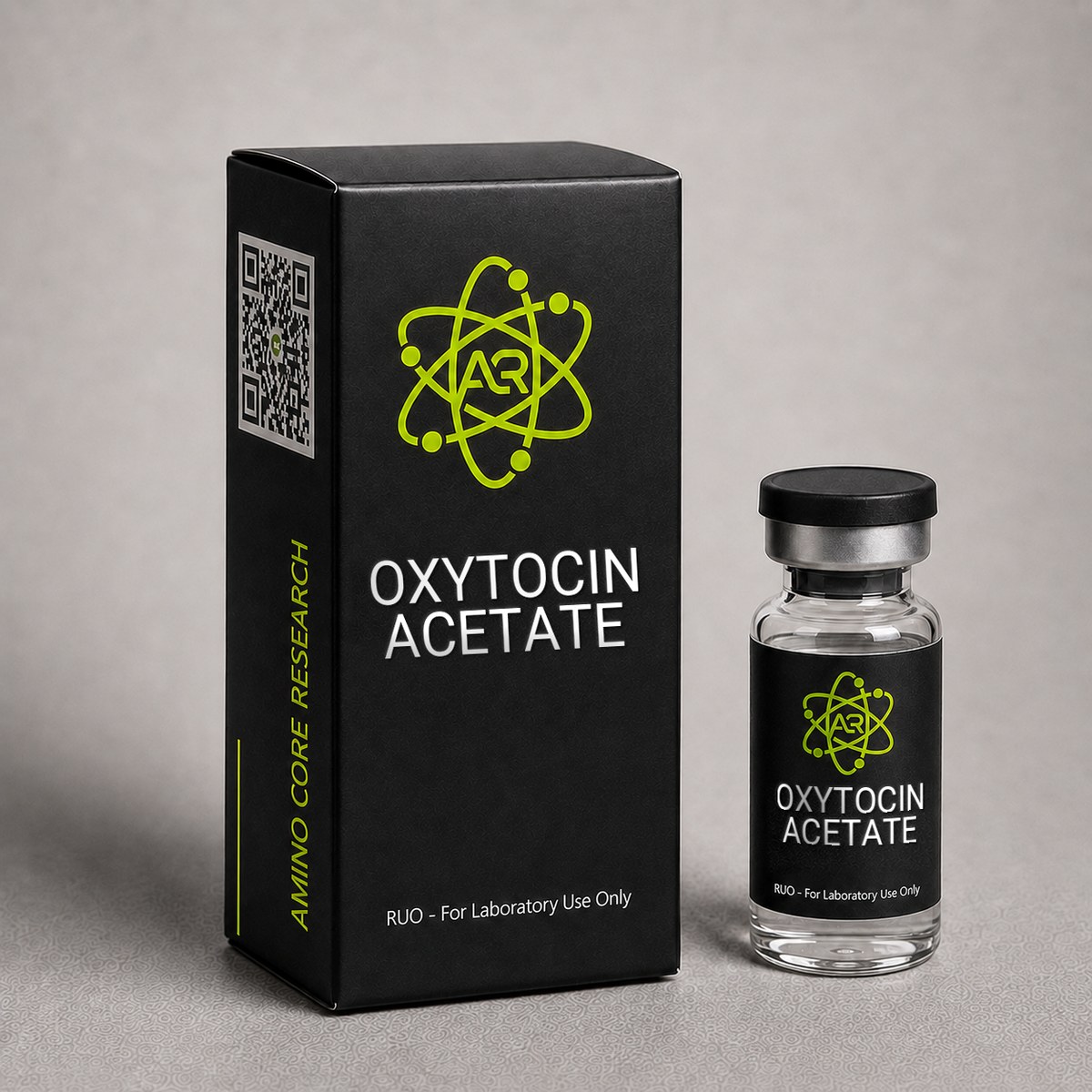
≥99%HPLC Purity
COAIncluded
USAShipped
Oxytocin Acetate Peptide
Cyclic nonapeptide hormone. Studied for oxytocin receptor signaling, social behavior neuroscience, and smooth muscle physiology research.
$35.00
In stock — ships next business day
Quick Facts
| SKU | ACR-OXY |
|---|---|
| CAS Number | 6233-83-6 |
| Molecular Formula | C43H66N12O12S2 |
| Molecular Weight | 1007.19 g/mol |
| Sequence | Cys-Tyr-Ile-Gln-Asn-Cys-Pro-Leu-Gly-NH2 |
| Purity | ≥99% |
| Physical Form | Lyophilized Powder |
| Storage | Store at -20°C |
What is Oxytocin?
Oxytocin is a cyclic nonapeptide hormone (Cys-Tyr-Ile-Gln-Asn-Cys-Pro-Leu-Gly-NH2) with a disulfide bridge between Cys1 and Cys6. First synthesized by Vincent du Vigneaud in 1953 (earning the Nobel Prize), it is produced in the hypothalamic paraventricular and supraoptic nuclei.
Oxytocin signals through the OXTR (oxytocin receptor), a Gq/11-coupled GPCR that activates phospholipase C, IP3-mediated calcium release, and downstream PKC signaling. Published research spans reproductive physiology, social neuroscience, cardiovascular function, and stress response modulation.
For laboratory research use only.
Mechanism of Action
Oxytocin (OXT) is a cyclic nonapeptide hormone with the sequence Cys-Tyr-Ile-Gln-Asn-Cys-Pro-Leu-Gly-NH2, featuring a disulfide bridge between Cys1 and Cys6 that creates a 20-membered ring with a C-terminal tripeptide tail. The molecule signals through a single G-protein-coupled receptor, the oxytocin receptor (OXTR), encoded by the OXTR gene on chromosome 3p25. OXTR exhibits sub-nanomolar to low-nanomolar affinity for oxytocin, with reported Ki values in the range of 0.5-2 nM in transfected human cell systems.[1]
Gq/11 Signaling — Primary Pathway
OXTR predominantly couples to Gq/11, activating phospholipase C-β (PLCβ), which hydrolyzes phosphatidylinositol 4,5-bisphosphate (PIP2) into inositol trisphosphate (IP3) and diacylglycerol (DAG). IP3 mobilizes intracellular Ca2+ from endoplasmic reticulum stores, while DAG activates protein kinase C (PKC). In myometrial smooth muscle, this Ca2+ surge drives myosin light-chain kinase (MLCK) activation and uterine contraction. In mammary myoepithelial cells, the same cascade triggers milk ejection.
Gi/o Signaling — Secondary Pathway
At higher receptor densities or under specific cellular contexts, OXTR can couple to Gi/o, inhibiting adenylyl cyclase and reducing cAMP. This pathway has been implicated in modulating neuronal excitability and presynaptic neurotransmitter release in central oxytocin circuits, including the hypothalamic paraventricular nucleus (PVN) and supraoptic nucleus (SON).
Central Nervous System Signaling
Within the CNS, oxytocin is synthesized in magnocellular and parvocellular neurons of the PVN and SON and is released both peripherally via the posterior pituitary and centrally via somatodendritic and axonal projections to limbic structures including the amygdala, nucleus accumbens, hippocampus, and prefrontal cortex. OXTR activation in the central amygdala has been shown in preclinical models to attenuate fear responses, while accumbal OXTR-dopamine D2 receptor heterodimer formation modulates social reward processing.[2]
Cross-Reactivity with Vasopressin Receptors
Oxytocin shares structural homology with arginine vasopressin (AVP), differing by only two residues (Ile3 vs Phe3; Leu8 vs Arg8). As a consequence, oxytocin exhibits measurable affinity for the three vasopressin receptor subtypes (V1aR, V1bR, V2R), with Ki values typically 10-100-fold higher than at OXTR. This cross-reactivity is an important confounder in interpreting research data, particularly at supraphysiological concentrations, and has driven development of selective OXTR agonists such as carbetocin and TGOT ([Thr4,Gly7]oxytocin).
Receptor Desensitization
OXTR undergoes rapid agonist-induced phosphorylation by G-protein-coupled receptor kinases (GRKs), recruitment of β-arrestin-1/2, internalization via clathrin-coated pits, and either recycling to the membrane or lysosomal degradation. This desensitization profile is relevant to research models examining tachyphylaxis during sustained oxytocin exposure.
Research & Clinical Studies
Intranasal Oxytocin and Social Cognition: Trust Game Study
One of the most widely cited investigations of oxytocin's effects on human social behavior was published by Kosfeld and colleagues in Nature (2005). This randomized, double-blind, placebo-controlled study examined whether intranasal oxytocin administration modulated interpersonal trust in healthy male participants engaged in an economic trust game.
Study Design
- Subjects: 128 healthy male university students
- Design: Double-blind, placebo-controlled, between-subjects
- Intervention: 24 IU intranasal oxytocin or placebo, administered 50 minutes prior to task
- Paradigm: Multi-round trust game with monetary stakes; "investors" allocated funds to anonymous "trustees"
- Control task: Risk experiment with non-social random payoff to isolate social vs general risk-taking effects
Key Results
- Investors receiving oxytocin transferred significantly more monetary units to trustees: median transfer of 9 vs 8 monetary units (placebo), p = 0.03
- 45% of oxytocin-treated investors displayed maximal trust (transferring all 12 units) vs 21% of placebo subjects
- No effect of oxytocin was observed in the non-social risk control condition, indicating the effect was specific to social risk-taking rather than general risk tolerance
- Oxytocin did not alter beliefs about trustee trustworthiness, suggesting the effect operates on behavioral approach rather than cognitive expectation
Research Context
This study catalyzed over a decade of subsequent research into central oxytocin effects on social cognition, fear processing, and pair bonding. Importantly, more recent meta-analyses (Nave et al., 2015; Declerck et al., 2020) have reported smaller effect sizes than the original, with publication bias and methodological heterogeneity contributing to ongoing debate about replicability. The Kosfeld study nonetheless established the 24 IU intranasal dose as a standard reference protocol in human oxytocin neuroscience research and remains a foundational citation in the field.[1]
Pharmacokinetic studies have since demonstrated that intranasal administration produces measurable elevations in cerebrospinal fluid oxytocin within 30-75 minutes, supporting the temporal design used in the trust game paradigm, though the proportion of administered peptide reaching central compartments remains a subject of ongoing investigation.
[1] Kosfeld M, Heinrichs M, Zak PJ, Fischbacher U, Fehr E. Oxytocin increases trust in humans. Nature. 2005;435(7042):673-676. PubMed ↗
Intranasal Oxytocin and Autism Spectrum Disorder: Social Function Trials
Oxytocin's role in social cognition has prompted extensive investigation into its potential as a research tool for understanding social deficits associated with autism spectrum disorder (ASD). Multiple randomized controlled trials have examined whether intranasal oxytocin administration modulates social behavior, eye gaze, and emotion recognition in individuals with ASD compared to neurotypical controls.
Landmark Trial Design
A pivotal multi-center randomized, double-blind, placebo-controlled trial published in Molecular Psychiatry (Yamasue et al., 2020) enrolled 106 adult male participants with high-functioning ASD across four sites in Japan. Participants received 48 IU of intranasal oxytocin daily (24 IU twice daily) or placebo for 6 weeks. The primary endpoint was the Autism Diagnostic Observation Schedule (ADOS) reciprocal social interaction score.
Key Findings
- ADOS reciprocity score improved significantly in the oxytocin group at week 2 and week 4, though the effect attenuated by week 6 (p < 0.05).
- Repetitive behavior subscale showed a statistically significant reduction (effect size d = 0.27).
- Plasma oxytocin concentrations correlated with behavioral response, suggesting dose-dependent pharmacodynamic effects.
- No significant change was observed in the primary social interaction endpoint at the 6-week timepoint, raising questions about tolerance development.
Functional Neuroimaging Correlates
Companion fMRI sub-studies demonstrated that intranasal oxytocin enhanced activity in the medial prefrontal cortex (mPFC) and anterior cingulate cortex (ACC) during social judgment tasks. These regions are part of the brain's default mode network and are consistently hypoactivated in ASD populations during social processing tasks. The amygdala showed task-dependent modulation, with decreased reactivity to threatening faces and increased reactivity to positive social cues.
Receptor Genotype Effects
Subgroup analyses revealed that participants carrying specific OXTR single nucleotide polymorphisms (SNPs), particularly rs53576 and rs2254298, demonstrated differential responses. GG homozygotes at rs53576 showed greater behavioral improvement than A-allele carriers, suggesting that endogenous receptor function modulates exogenous oxytocin responsiveness. This pharmacogenomic finding has implications for designing future research stratification protocols.
Tachyphylaxis and Dosing Considerations
The attenuation of effects over the 6-week treatment period suggests possible receptor desensitization or downregulation with chronic dosing. Pharmacokinetic modeling indicates that intermittent or pulsatile dosing schedules may preserve receptor sensitivity better than continuous daily administration. This finding parallels observations in other GPCR systems where chronic agonist exposure produces internalization via β-arrestin-mediated mechanisms.
Research Implications
The Yamasue trial and subsequent meta-analyses (including the SOARS-B trial in pediatric ASD) have refined the field's understanding of oxytocin's translational potential. Current research focuses on identifying responder subpopulations, optimizing dosing regimens to avoid tachyphylaxis, and developing oxytocin receptor-selective analogs that may overcome the pharmacokinetic limitations of native oxytocin. These investigations remain strictly preclinical and exploratory in laboratory research contexts.
[1] Yamasue H, Okada T, Munesue T, et al. Effect of intranasal oxytocin on the core social symptoms of autism spectrum disorder: a randomized clinical trial. Molecular Psychiatry. 2020;25(8):1849-1858. PubMed ↗
[2] Sikich L, Kolevzon A, King BH, et al. Intranasal Oxytocin in Children and Adolescents with Autism Spectrum Disorder. New England Journal of Medicine. 2021;385(16):1462-1473. PubMed ↗
Chemical & Physical Properties
The following table summarizes verified physicochemical data for oxytocin acetate as cataloged in PubChem and standard pharmacopoeial references. Values are provided for the free peptide; the acetate salt form has slightly higher mass due to associated acetate counterions but identical primary sequence and bioactivity.
| Full Name | Oxytocin (free peptide); supplied as Oxytocin Acetate salt |
|---|---|
| Synonyms | OXT, Pitocin, Syntocinon, α-Hypophamine, Ocytocin |
| Molecular Formula | C43H66N12O12S2 (free peptide) |
| Molecular Weight | 1,007.19 g/mol (free peptide) |
| CAS Number | 50-56-6 (free peptide) / 6233-83-6 (acetate salt) |
| Sequence | H-Cys-Tyr-Ile-Gln-Asn-Cys-Pro-Leu-Gly-NH2 |
| One-Letter Code | CYIQNCPLG-NH2 (disulfide Cys1-Cys6) |
| Amino Acid Count | 9 residues (nonapeptide) |
| Origin / Developer | Isolated by Vincent du Vigneaud (Nobel Prize, Chemistry, 1955) — first peptide hormone to be sequenced and chemically synthesized |
| Key Modifications | Intramolecular disulfide bridge (Cys1-Cys6) forming a 20-membered cyclic ring; C-terminal glycinamide (-NH2) |
| Physical Form | White to off-white lyophilized powder |
| Solubility | Soluble in water (≥1 mg/mL), sterile bacteriostatic water, dilute acetic acid; sparingly soluble in alcohols |
| Purity | ≥99% (HPLC) |
| Isoelectric Point (pI) | ~7.7 |
| Net Charge at pH 7 | +1 (free amine at N-terminus) |
| UniProt (Precursor) | P01178 (OXT_HUMAN, oxytocin-neurophysin 1 preproprotein) |
Oxytocin is one of the smallest and most thoroughly characterized peptide hormones in pharmacology, and its synthesis by du Vigneaud in 1953 represented the first chemical synthesis of a polypeptide hormone. The disulfide bridge is essential for receptor binding affinity — reduction of the Cys1-Cys6 bond abolishes activity at OXTR.
Handling & Reconstitution Guidelines
Oxytocin acetate is supplied as a lyophilized powder and requires careful reconstitution to preserve peptide integrity. The cyclic disulfide bridge between Cys1 and Cys6 is essential for receptor binding activity, and improper handling can lead to disulfide scrambling, oxidation, or aggregation that compromises bioactivity.
Reconstitution Protocol
- Allow the vial to reach room temperature before opening. Remove from -20°C storage and let stand at ambient temperature for 15-20 minutes to prevent moisture condensation on the lyophilized powder.
- Select the appropriate diluent: bacteriostatic water (0.9% benzyl alcohol) is preferred for multi-use reconstitution, while sterile water for injection or 0.9% sodium chloride is suitable for single-use applications.
- Calculate target concentration: For a 5 mg vial, add 5 mL of diluent to achieve a 1 mg/mL working solution. For higher concentrations, use 2.5 mL to obtain 2 mg/mL.
- Inject diluent slowly down the side of the vial wall, avoiding direct impact on the lyophilized cake. Do not inject directly onto the powder.
- Swirl gently in a circular motion until fully dissolved. Do not shake or vortex — agitation can denature the peptide and disrupt the disulfide bridge.
- Inspect visually for complete dissolution. The solution should be clear and colorless. Any cloudiness, particulates, or discoloration indicates degradation and the vial should be discarded.
Compound-Specific Handling Notes
Disulfide bridge sensitivity: Oxytocin contains a single intramolecular disulfide bond (Cys1-Cys6) that is critical for the cyclic conformation required for OXTR binding. Exposure to reducing agents (DTT, β-mercaptoethanol, TCEP) will cleave this bridge and abolish activity. Avoid contact with metal surfaces that may catalyze oxidation.
pH considerations: Oxytocin is most stable at slightly acidic pH (3.5-4.5). Avoid alkaline buffers, which accelerate degradation via deamidation of Asn5 and Gln4 residues.
Adsorption losses: Oxytocin exhibits non-specific binding to glass and plastic surfaces, particularly at low concentrations (<10 μg/mL). For dilute working solutions, consider adding 0.1% bovine serum albumin (BSA) as a carrier protein to minimize adsorption losses.
Temperature sensitivity: Once reconstituted, maintain the solution at 2-8°C and avoid repeated warming. Use cold-chain protocols for any solution transfers.
Aliquoting Recommendations
For long-term research use, prepare single-use aliquots immediately after reconstitution to avoid freeze-thaw cycles. Use low-binding polypropylene tubes (Eppendorf Protein LoBind or equivalent) and store at -80°C for maximum stability. Each freeze-thaw cycle can reduce potency by approximately 5-10%.
Storage & Stability Information
Proper storage of oxytocin acetate is essential for maintaining peptide integrity and reproducible research results. The compound's stability profile is dictated by its cyclic disulfide structure, susceptibility to deamidation, and sensitivity to oxidative degradation.
Lyophilized Powder Storage
- Long-term storage (>1 month): Store at -20°C in original sealed vial with desiccant. Under these conditions, lyophilized oxytocin acetate remains stable for 24-36 months with minimal loss of potency.
- Extended long-term storage: For storage exceeding 2 years, -80°C is recommended to minimize gradual degradation pathways including aspartimide formation and disulfide scrambling.
- Short-term storage (<30 days): Stable at 2-8°C (refrigerated) when sealed and protected from moisture.
- Transit conditions: Oxytocin acetate tolerates ambient temperatures (15-25°C) for up to 7 days during shipping without significant degradation, provided the vial remains sealed.
Reconstituted Solution Stability
- Refrigerated (2-8°C): Reconstituted solutions in bacteriostatic water remain stable for 14-21 days. Solutions in sterile water without preservative should be used within 7 days.
- Frozen aliquots (-20°C or -80°C): Stable for 3-6 months in low-binding polypropylene tubes. Limit to a maximum of 2-3 freeze-thaw cycles.
- Room temperature: Reconstituted oxytocin degrades rapidly at ambient temperature, with measurable potency loss within 24-48 hours. Avoid prolonged room temperature exposure.
Degradation Pathways
Understanding oxytocin's degradation chemistry helps researchers anticipate stability issues:
- Disulfide reduction/scrambling: The Cys1-Cys6 bridge can be reduced under anaerobic conditions or scrambled in the presence of free thiols, producing inactive linear or misfolded species.
- Deamidation: Asn5 and Gln4 are susceptible to deamidation, particularly at pH >6, producing iso-aspartate and glutamate derivatives with reduced OXTR affinity.
- Oxidation: Although oxytocin lacks methionine, the disulfide bridge can undergo oxidation to sulfoxide or sulfone forms under exposure to peroxides or transition metals.
- Aggregation: At high concentrations or low pH, oxytocin can form fibrillar aggregates, particularly when subjected to mechanical stress (shaking, vortexing).
Best Practices Summary
For optimal research reproducibility: store lyophilized material at -20°C with desiccant, reconstitute only what is needed for immediate experiments, prepare single-use aliquots to avoid freeze-thaw degradation, protect from light using amber vials or foil wrapping, and verify peptide integrity periodically via HPLC or mass spectrometry for long-stored material. Always document reconstitution dates and storage history for each experimental batch.
Frequently Asked Questions
What is Oxytocin?
Oxytocin is a cyclic 9-amino acid hormone with a Cys1-Cys6 disulfide bridge. It signals through the OXTR receptor via Gq/PLC/calcium pathways. Research spans neuroscience, reproductive biology, and behavioral pharmacology. For research use only.
What is the molecular weight and CAS number of Oxytocin Acetate?
Oxytocin has a molecular formula of C43H66N12O12S2 and a molecular weight of 1,007.19 g/mol as the free peptide. The CAS number for free oxytocin is 50-56-6, while the acetate salt form is registered under CAS 6233-83-6. It is a cyclic nonapeptide containing nine amino acid residues (Cys-Tyr-Ile-Gln-Asn-Cys-Pro-Leu-Gly-NH2) with an intramolecular disulfide bridge between Cys1 and Cys6 and a C-terminal glycinamide. Purity supplied for research use is ≥99% by HPLC.
How does Oxytocin differ from Vasopressin?
Oxytocin and arginine vasopressin (AVP) are structural homologs that differ by only two amino acid residues: oxytocin contains isoleucine at position 3 and leucine at position 8, while vasopressin has phenylalanine and arginine, respectively. Both are cyclic nonapeptides with a Cys1-Cys6 disulfide bridge. Despite this similarity, they activate distinct receptor systems — oxytocin preferentially binds the oxytocin receptor (OXTR), while vasopressin acts on V1a, V1b, and V2 receptors. Functional cross-reactivity exists at supraphysiological concentrations. Research applications differ accordingly: oxytocin is studied in social cognition, parturition, and lactation models, while vasopressin is studied in osmoregulation and blood pressure research.
How should Oxytocin Acetate be stored?
Lyophilized oxytocin acetate should be stored at -20°C for long-term stability (>12 months), at 2-8°C for short-term storage (up to several weeks), and may be shipped at ambient temperature for brief transit periods. After reconstitution in sterile or bacteriostatic water, the solution should be stored at 2-8°C and used within 7-14 days to minimize peptide degradation. The disulfide bridge is susceptible to reduction and oxidative cleavage, so repeated freeze-thaw cycles, exposure to reducing agents, and prolonged room-temperature storage should be avoided. Aliquoting reconstituted material into single-use volumes is recommended for research workflows.
Does Oxytocin require a disulfide bridge for activity?
Yes, the intramolecular disulfide bridge between Cys1 and Cys6 is essential for oxytocin's biological activity. This bond creates a 20-membered cyclic ring that constrains the peptide into the conformation required for high-affinity binding at the oxytocin receptor (OXTR). Reduction of the disulfide bond — for example, by exposure to dithiothreitol (DTT), β-mercaptoethanol, or strongly reducing cellular environments — produces a linear nonapeptide that is essentially inactive at OXTR. This structural requirement is shared with the related peptide vasopressin and has guided the design of synthetic analogs such as carbetocin, in which the disulfide is replaced by a more stable carba-bridge to prolong half-life.
What sizes of Oxytocin Acetate are available from AminoCore Research?
AminoCore Research typically offers Oxytocin Acetate in multiple vial sizes to accommodate different research applications, commonly including 2 mg, 5 mg, and 10 mg lyophilized powder presentations. All vials are supplied with ≥99% HPLC purity verification and a certificate of analysis (COA). Larger bulk quantities may be available upon request for institutional research programs. All material is sold strictly for laboratory research use and is not intended for human or veterinary therapeutic application.
Does Oxytocin Acetate cross the blood-brain barrier in research models?
Native oxytocin exhibits limited blood-brain barrier (BBB) penetration when administered peripherally, with less than 1-2% of circulating peptide reaching cerebrospinal fluid under normal conditions. Research investigating central oxytocin effects typically employs intranasal administration, which provides nose-to-brain transport via the olfactory and trigeminal pathways, bypassing the BBB. Studies have demonstrated measurable CSF oxytocin elevation within 30-60 minutes following intranasal dosing. Intracerebroventricular (ICV) administration is also used in preclinical rodent models to bypass BBB limitations and directly assess central receptor activation.
What is the half-life of Oxytocin in research applications?
Oxytocin has a notably short plasma half-life of approximately 3-6 minutes in humans and rodent models, primarily due to rapid enzymatic degradation by oxytocinase (cystyl aminopeptidase) and clearance by the liver and kidneys. This brief pharmacokinetic profile necessitates continuous infusion or repeated dosing in many research protocols. Central nervous system effects following intranasal administration may persist longer (30-90 minutes) due to slower CSF clearance. The short half-life has motivated development of oxytocin receptor agonists with extended pharmacokinetic properties, such as carbetocin, for comparative research.
How does Oxytocin Acetate compare to Carbetocin in research applications?
Both compounds activate the oxytocin receptor (OXTR), but they differ substantially in pharmacokinetic and structural properties. Carbetocin is a synthetic long-acting analog with the disulfide bridge replaced by a thioether (monocarba) modification and N-terminal deamination, conferring resistance to oxytocinase degradation and extending plasma half-life to approximately 40 minutes versus 3-6 minutes for native oxytocin. Carbetocin shows higher selectivity for OXTR over vasopressin receptors, while oxytocin has measurable cross-reactivity with V1a receptors. Oxytocin remains the gold-standard reference compound for OXTR research due to its endogenous status and extensively characterized signaling profile.
For laboratory and research use only. This product is not for human or veterinary use. It is not a drug, supplement or food.
This product is not FDA approved for any indication, and is not intended to diagnose, treat, cure, or prevent any disease. All product information is derived from published preclinical research and does not constitute medical advice or claims.