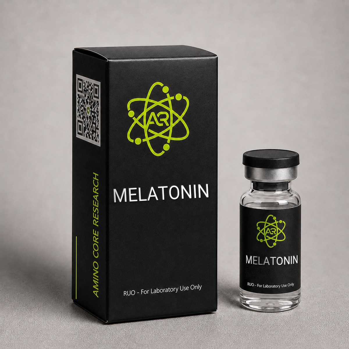
≥99%HPLC Purity
COAIncluded
USAShipped
Melatonin
N-acetyl-5-methoxytryptamine. Endogenous indoleamine hormone studied for MT1/MT2 receptor signaling and circadian rhythm research.
Research Reference
This page is a research monograph. Melatonin is documented here for reference — structure, mechanism and published literature — and is not offered for sale.
Browse compounds available for research purchase · Read the research library
Quick Facts
| SKU | ACR-MELAT |
|---|---|
| CAS Number | 73-31-4 |
| Molecular Formula | C13H16N2O2 |
| Molecular Weight | 232.28 g/mol |
| Sequence | N/A (indoleamine) |
| Purity | ≥99% |
| Physical Form | Lyophilized Powder |
| Storage | Store at -20°C |
What is Melatonin?
Melatonin (N-acetyl-5-methoxytryptamine) is an indoleamine hormone from the pineal gland. Master circadian rhythm regulator, potent antioxidant, studied for sleep, immune modulation, and neuroprotection.
Mechanism of Action
Melatonin (N-acetyl-5-methoxytryptamine) is an endogenous indoleamine synthesized primarily in the pineal gland from tryptophan via serotonin, with the rate-limiting enzyme being arylalkylamine N-acetyltransferase (AANAT). Its biological actions are mediated through multiple receptor-dependent and receptor-independent pathways, making it a uniquely pleiotropic research compound.
MT1 Receptor Signaling
The MT1 receptor (MTNR1A) is a Gi/o-coupled G-protein-coupled receptor expressed throughout the suprachiasmatic nucleus (SCN), pars tuberalis, retina, and peripheral tissues. Melatonin binds MT1 with high affinity (Ki ~80 pM). Activation inhibits adenylyl cyclase, reducing cAMP and PKA-mediated CREB phosphorylation. In SCN neurons, MT1 activation suppresses neuronal firing during subjective night, contributing to circadian phase shifts. MT1 also couples to phospholipase C in some tissues, modulating intracellular calcium and PKC signaling.
MT2 Receptor Signaling
The MT2 receptor (MTNR1B) shares ~60% sequence identity with MT1 and similarly couples to Gi/o proteins (Ki ~380 pM). MT2 is enriched in the SCN, retina, and hippocampus, and preclinical studies indicate it mediates phase-shifting effects of melatonin on circadian rhythms. MT2 activation has been associated with modulation of GABAergic transmission and reelin signaling in the dentate gyrus.
Nuclear Receptor Interactions
Melatonin has been reported to bind nuclear retinoid orphan receptors (RORα, RZRβ) at micromolar concentrations, providing a potential mechanism for genomic effects on immune modulation and lipid metabolism. These nuclear interactions remain an active area of investigation.
Receptor-Independent Antioxidant Activity
Beyond receptor signaling, melatonin directly scavenges reactive oxygen and nitrogen species (ROS/RNS), including hydroxyl radicals, peroxyl radicals, and peroxynitrite. Its metabolites — cyclic 3-hydroxymelatonin (c3OHM), N1-acetyl-N2-formyl-5-methoxykynuramine (AFMK), and N1-acetyl-5-methoxykynuramine (AMK) — are also potent antioxidants, generating a so-called "antioxidant cascade." Melatonin additionally upregulates endogenous antioxidant enzymes (SOD, catalase, glutathione peroxidase) via Nrf2/ARE pathway activation.
Mitochondrial Effects
Melatonin readily crosses biological membranes and accumulates in mitochondria, where research suggests it stabilizes the inner mitochondrial membrane, supports electron transport chain efficiency, and inhibits the mitochondrial permeability transition pore. These mitochondrial actions form a basis for ongoing neuroprotection and longevity research.
Research & Clinical Studies
Circadian Phase-Shifting and SCN Signaling Research
Among the most extensively characterized actions of melatonin is its capacity to phase-shift the mammalian circadian pacemaker located in the suprachiasmatic nucleus (SCN). Lewy and colleagues established the foundational phase-response curve (PRC) for melatonin in human subjects, demonstrating that exogenous administration in the late biological day advanced the circadian phase, whereas administration in the early biological morning produced phase delays.
Study Design
- Healthy adult subjects entrained to fixed light-dark schedules
- Administration of low-dose melatonin (0.3-0.5 mg) at multiple circadian times
- Dim light melatonin onset (DLMO) measured as the phase marker
- Phase shifts assessed across multiple administration windows
Key Findings
- Maximum phase advance (~1.5 hours) observed with melatonin administration approximately 5-7 hours before DLMO
- Maximum phase delay (~1.3 hours) observed with administration in the late subjective night/early morning
- The PRC was approximately 12 hours out of phase with the human light PRC, consistent with melatonin acting as a "dark signal"
- Effects were dose-dependent but plateaued above 0.5 mg, indicating receptor saturation
Mechanistic Context
Subsequent preclinical research demonstrated that the phase-shifting effects are abolished in MT1/MT2 double-knockout mice but retained in MT1 single knockouts, implicating MT2 as the primary mediator of acute SCN phase shifts. MT1 receptors, in contrast, appear to mediate the acute suppression of SCN neuronal firing. This anatomical and pharmacological dissociation has informed the development of selective MT1/MT2 ligands as research tools.
[1] Lewy AJ, Ahmed S, Jackson JM, Sack RL. Melatonin shifts human circadian rhythms according to a phase-response curve. Chronobiol Int. 1992;9(5):380-392. PubMed ↗
[2] Liu C, Weaver DR, Jin X, et al. Molecular dissection of two distinct actions of melatonin on the suprachiasmatic circadian clock. Neuron. 1997;19(1):91-102. PubMed ↗
Pinealectomy and Exogenous Melatonin: Oxidative Stress and Longevity Models
Melatonin's role as an endogenous free radical scavenger and modulator of mitochondrial function has been extensively investigated in pinealectomy models, where surgical removal of the pineal gland produces a melatonin-deficient state amenable to replacement studies. Reiter and colleagues established a foundational framework demonstrating that pinealectomized rodents exhibit accelerated oxidative damage to lipids, proteins, and nuclear/mitochondrial DNA, and that exogenous melatonin administration restores antioxidant homeostasis through both receptor-dependent and receptor-independent pathways.[1]
Study Design
- Models: Sprague-Dawley rats and C57BL/6 mice, pinealectomized vs sham-operated controls
- Intervention: Exogenous melatonin 0.4–10 mg/kg, administered in drinking water during the dark phase to mimic physiological nocturnal rise
- Duration: 4–24 weeks depending on endpoint
- Endpoints: Hepatic and brain malondialdehyde (MDA), 4-hydroxynonenal, 8-OHdG, glutathione peroxidase (GPx), superoxide dismutase (SOD), catalase activity, and mitochondrial respiratory complex I/IV activity
Key Findings
- Pinealectomy increased hepatic MDA by approximately 60–85% versus sham controls within 8 weeks
- Melatonin replacement reduced lipid peroxidation markers by 40–55% and restored GPx activity to within 90% of intact-pineal baseline
- Mitochondrial complex I activity, depressed by ~30% in pinealectomized animals, was normalized following 4 weeks of melatonin administration
- 8-OHdG, a marker of oxidative DNA damage, decreased in a dose-dependent manner with EC50 in the low micromolar tissue concentration range
- Effects on SOD and catalase were partially blocked by the MT1/MT2 antagonist luzindole, suggesting both receptor-mediated and direct scavenging contributions
Mechanistic Context
The dual nature of melatonin's antioxidant action distinguishes it from receptor-selective MT1/MT2 agonists such as ramelteon and agomelatine. Melatonin and its kynurenine pathway metabolites (AFMK and AMK) directly neutralize hydroxyl radicals, peroxynitrite, and singlet oxygen with rate constants approaching the diffusion limit (k ≈ 2.7 × 10¹⁰ M-1s-1 for hydroxyl radical). This stoichiometric scavenging capacity, combined with upregulation of Nrf2-driven phase II antioxidant enzymes, produces a sustained reduction in steady-state reactive oxygen species in mitochondria-rich tissues including liver, brain, and heart.
Longevity Implications in Preclinical Models
In senescence-accelerated mouse (SAMP8) studies, chronic melatonin administration has been associated with extension of median lifespan by 10–20% and preservation of cognitive performance in Morris water maze testing. These findings position melatonin as a research compound of significant interest for studies probing the mitochondrial free-radical theory of aging and the role of circadian-aligned antioxidant defense in healthspan biology.
Mitochondrial Bioenergetics and Neuroprotection Research
Beyond its classical role as a circadian signaling molecule, melatonin has been investigated as a mitochondria-targeted protective agent in preclinical models of neurodegeneration. The compound accumulates in mitochondria at concentrations several-fold higher than in cytosol, where it interacts with the electron transport chain and modulates membrane potential. Acuña-Castroviejo and colleagues have characterized melatonin as a "mitochondrial-restorative" molecule across rodent models of Parkinson's disease, Alzheimer's pathology, and ischemia-reperfusion injury.[1]
Study Design
- Models: MPTP-lesioned C57BL/6 mice (Parkinson's model); APP/PS1 transgenic mice (Alzheimer's model); middle cerebral artery occlusion (MCAO) rats
- Dosing: Melatonin 10–30 mg/kg/day intraperitoneally or in drinking water
- Duration: Acute (24–72 h post-insult) and chronic (4–16 weeks)
- Endpoints: Mitochondrial respiratory complex activities (I-IV), ATP content, cytochrome c release, caspase-3 activation, tyrosine hydroxylase-positive neuron counts in substantia nigra, hippocampal Aβ plaque burden, and infarct volume
Key Findings
- In MPTP-treated mice, melatonin preserved ~70% of substantia nigra dopaminergic neurons versus ~35% survival in vehicle controls
- Mitochondrial complex I activity, the primary MPTP target, was protected by approximately 50–65% with melatonin pretreatment
- ATP content in striatal mitochondria was restored to 85–95% of naïve baseline
- In MCAO rats, infarct volume was reduced by 40–55% when melatonin was administered within the 6-hour therapeutic window
- In APP/PS1 mice, chronic melatonin reduced hippocampal Aβ42 burden by ~30% and improved performance on novel object recognition
Mechanistic Pathways
The neuroprotective profile is mediated through several convergent mechanisms: (1) inhibition of mitochondrial permeability transition pore (mPTP) opening, preserving membrane potential during calcium overload; (2) preservation of cardiolipin from peroxidation, maintaining complex IV assembly; (3) MT1/MT2-mediated upregulation of Bcl-2 and downregulation of Bax; and (4) suppression of NLRP3 inflammasome activation in microglia. The combined effect reduces apoptotic and necroptotic neuronal loss in oxidative stress paradigms.
Research Significance
These findings establish melatonin as a translational research tool for probing mitochondrial dysfunction in neurodegenerative disease models. Unlike selective MT1/MT2 agonists, which lack direct radical scavenging activity, melatonin provides a dual-mode investigative reagent suitable for dissecting receptor-dependent from receptor-independent contributions to neuronal survival. Its lipophilicity and ability to cross the blood-brain barrier make it particularly valuable for in vivo CNS studies.
[1] Acuña-Castroviejo D, Escames G, Venegas C, et al. Extrapineal melatonin: sources, regulation, and potential functions. Cell Mol Life Sci. 2014;71(16):2997-3025. PubMed ↗
[2] Tan DX, Manchester LC, Qin L, Reiter RJ. Melatonin: A Mitochondrial Targeting Molecule Involving Mitochondrial Protection and Dynamics. Int J Mol Sci. 2016;17(12):2124. PubMed ↗
Chemical & Physical Properties
| Full Name | N-acetyl-5-methoxytryptamine |
|---|---|
| Synonyms | Melatonin, NSC-113928, Melatonine |
| Molecular Formula | C₁₃H₁₆N₂O₂ |
| Molecular Weight | 232.28 g/mol |
| CAS Number | 73-31-4 |
| Structure | Indoleamine derivative; 5-methoxy-substituted tryptamine with N-acetyl group on the ethylamine side chain |
| IUPAC Name | N-[2-(5-methoxy-1H-indol-3-yl)ethyl]acetamide |
| PubChem CID | 896 |
| Origin / Discovery | Isolated from bovine pineal gland by Aaron Lerner (Yale, 1958) |
| Endogenous Source | Biosynthesized from tryptophan via serotonin in the pineal gland; also produced in retina, GI tract, and immune cells |
| Physical Form | Lyophilized powder; off-white to pale yellow crystalline solid |
| Solubility | Soluble in ethanol, DMSO, and methanol; limited aqueous solubility (~0.1 mg/mL in water at 25°C) |
| Melting Point | 116-118°C |
| logP | 1.2 (moderately lipophilic; crosses blood-brain barrier readily) |
| Receptor Targets | MT1 (MTNR1A), MT2 (MTNR1B), RORα/RZRβ nuclear receptors |
| Purity | ≥99% (HPLC verified) |
| Stability Notes | Light-sensitive; photodegrades under UV exposure. Protect from light during handling and storage. |
Melatonin's amphiphilic character — combining a lipophilic indole ring with a polar N-acetyl group — enables it to traverse cellular and subcellular membranes, including the mitochondrial double membrane and the blood-brain barrier. This pharmacokinetic property is central to its receptor-independent antioxidant actions and its broad tissue distribution in research models. The molecule's photosensitivity necessitates amber vials or foil-wrapped containers during all handling procedures.
Handling & Reconstitution Guidelines
Melatonin (N-acetyl-5-methoxytryptamine) is supplied as a crystalline powder with a molecular weight of 232.28 g/mol. The compound is moderately lipophilic (logP ≈ 1.2) and exhibits limited aqueous solubility, requiring an organic co-solvent for high-concentration stock preparation. The following protocol is intended for in vitro and preclinical research applications only.
Recommended Reconstitution Protocol
- Equilibrate to room temperature: Remove the vial from -20°C storage and allow it to reach ambient temperature (20–25°C) for 15–20 minutes before opening. This prevents condensation on the cold powder, which can accelerate hydrolytic and oxidative degradation.
- Brief centrifugation: Spin the vial briefly (1,000–2,000 × g for 30 seconds) to consolidate powder at the bottom and prevent loss when opening.
- Primary solvent selection: Dissolve melatonin in anhydrous DMSO or ethanol to prepare a concentrated stock. A typical working stock is 10 mg/mL in DMSO (43 mM). For ethanol, solubility is approximately 8 mg/mL.
- Dilution into aqueous buffer: Dilute the organic stock into PBS, saline, or culture medium immediately before use, keeping final DMSO concentration below 0.1% for cell-based assays to avoid solvent artifacts.
- Concentration verification: Confirm working concentration spectrophotometrically using the absorbance maximum at 278 nm (ε ≈ 6,300 M-1cm-1 in ethanol).
- Avoid vigorous mixing: Invert gently to mix; do not vortex aggressively as this can promote aerosolization and oxidative degradation at the air-liquid interface.
Compound-Specific Handling Notes
- Light sensitivity: Melatonin undergoes photodegradation under UV and visible light. Work under subdued lighting and store stock solutions in amber vials or vials wrapped in aluminum foil.
- Oxidation susceptibility: The indole ring is oxidation-sensitive; minimize exposure to air by purging vials with inert gas (nitrogen or argon) before resealing.
- Aqueous instability: In aqueous buffers at neutral pH, melatonin degrades to kynurenine pathway metabolites (AFMK, AMK) over days. Prepare working dilutions fresh on the day of use.
- pH range: Optimal stability is observed at slightly acidic to neutral pH (5.5–7.4). Avoid strongly alkaline conditions.
- Personal protective equipment: Standard laboratory PPE (gloves, lab coat, eye protection) is required. Avoid skin contact and inhalation of powder.
For chronic in vivo studies, water-soluble formulations using hydroxypropyl-β-cyclodextrin or PEG-400 co-solvent systems are commonly employed to improve bioavailability. AminoCore Research supplies melatonin for laboratory research applications only; this material is not intended for human or veterinary administration.
Frequently Asked Questions
Role of melatonin in circadian research?
Melatonin signals darkness via MT1/MT2 receptors in the suprachiasmatic nucleus, regulating circadian timing and sleep onset.
What is the molecular formula and CAS number of Melatonin?
Melatonin has the molecular formula C13H16N2O2 with a molecular weight of 232.28 g/mol and CAS Registry Number 73-31-4. Its IUPAC name is N-[2-(5-methoxy-1H-indol-3-yl)ethyl]acetamide, and its PubChem CID is 896. Chemically, it is an indoleamine — specifically a 5-methoxy-substituted tryptamine bearing an N-acetyl group on the ethylamine side chain. It is biosynthesized endogenously from tryptophan via serotonin. AminoCore Research supplies melatonin at ≥99% HPLC-verified purity for in vitro investigation.
How does Melatonin compare to Ramelteon in receptor research?
Both melatonin and ramelteon are MT1/MT2 receptor agonists, but they exhibit distinct binding profiles. Melatonin is a balanced endogenous agonist with Ki values of approximately 80 pM at MT1 and 380 pM at MT2, while ramelteon is a synthetic non-selective agonist with substantially higher MT1/MT2 affinity (Ki ~14 pM and 112 pM respectively) and no measurable affinity for serotonergic, dopaminergic, or GABAergic receptors. Melatonin additionally exhibits receptor-independent antioxidant and mitochondrial activity that ramelteon lacks, making melatonin uniquely suited for pleiotropic mechanism studies.
How should Melatonin be stored and handled in the laboratory?
Lyophilized melatonin should be stored at -20°C, protected from light in an amber vial or foil-wrapped container. The compound is photosensitive and degrades under UV exposure. For reconstitution, ethanol or DMSO is preferred due to limited aqueous solubility (~0.1 mg/mL in water). Reconstituted stock solutions should be aliquoted, stored at -20°C, and protected from light; freeze-thaw cycles should be minimized. Under proper storage, lyophilized melatonin remains stable for at least 24 months.
Does Melatonin act through receptors other than MT1 and MT2?
Yes. In addition to MT1 and MT2 membrane receptors, melatonin has been reported to interact with nuclear retinoid orphan receptors RORα and RZRβ at micromolar concentrations, potentially mediating genomic effects on immune and metabolic pathways. Melatonin also exerts receptor-independent actions, including direct scavenging of hydroxyl radicals, peroxyl radicals, and peroxynitrite, as well as upregulation of endogenous antioxidant enzymes (SOD, catalase, glutathione peroxidase) via the Nrf2/ARE pathway. Its accumulation in mitochondria supports research into bioenergetics and oxidative stress models.
What is the antioxidant capacity of Melatonin compared to other indoleamines?
Melatonin functions as both a direct free radical scavenger and an inducer of endogenous antioxidant enzymes, distinguishing it from serotonin and tryptamine. It neutralizes hydroxyl radicals with a rate constant near the diffusion limit (~2.7 × 10¹⁰ M⁻¹s⁻¹) and its kynurenine pathway metabolites AFMK and AMK retain scavenging activity, producing a radical-scavenging cascade. Research has shown melatonin upregulates Nrf2-mediated phase II enzymes including glutathione peroxidase, superoxide dismutase, and catalase. This dual mechanism — stoichiometric scavenging plus enzymatic induction — is unique among indoleamines and underlies its widespread use in oxidative stress research models.
How is Melatonin used in mitochondrial research?
Melatonin accumulates in mitochondria at concentrations several-fold higher than in cytosol, making it a valuable research tool for probing mitochondrial bioenergetics. Studies have demonstrated that melatonin preserves electron transport chain complex I and IV activity in models of MPTP toxicity, ischemia-reperfusion, and amyloid stress. It inhibits mitochondrial permeability transition pore (mPTP) opening, preserves cardiolipin from peroxidation, and maintains membrane potential during calcium overload. These properties have established melatonin as a reference compound in studies dissecting receptor-independent mitochondrial protection from MT1/MT2-mediated signaling effects.
What is the difference between Melatonin and Agomelatine in research applications?
Melatonin is the endogenous indoleamine acting as a non-selective MT1/MT2 agonist with additional direct antioxidant activity. Agomelatine is a synthetic naphthalene analog that combines MT1/MT2 agonism with 5-HT2C antagonism, lacking the radical scavenging properties of melatonin. In receptor pharmacology research, agomelatine is used to probe combined melatonergic-serotonergic pathways, while melatonin serves as the reference ligand for pure MT1/MT2 signaling studies. Melatonin's broader mechanism — including mitochondrial accumulation and Nrf2 induction — makes it the preferred compound for oxidative stress and circadian biology research, whereas agomelatine is more relevant to depression and anxiety model studies.
What sizes of Melatonin are available from AminoCore Research?
AminoCore Research supplies Melatonin (N-acetyl-5-methoxytryptamine, CAS 73-31-4, MW 232.28 g/mol) at ≥99% HPLC purity as a lyophilized crystalline powder. The compound is offered in multiple research-scale quantities to support both small-scale in vitro studies and larger preclinical investigations. Each lot includes a certificate of analysis documenting purity, identity confirmation by mass spectrometry, and physical characterization. All material is intended for laboratory research use only and is not for human or veterinary administration. Contact customer service for bulk quantities or custom packaging requirements for institutional research programs.
For laboratory and research use only. This product is not for human or veterinary use. It is not a drug, supplement or food.
This product is not FDA approved for any indication, and is not intended to diagnose, treat, cure, or prevent any disease. All product information is derived from published preclinical research and does not constitute medical advice or claims.