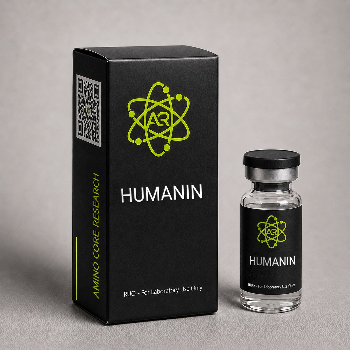
≥99%HPLC Purity
COAIncluded
USAShipped
Humanin Peptide
Mitochondria-derived peptide (MDP) researched for potent cytoprotective and anti-apoptotic properties. Studied in neurodegeneration, Alzheimer disease models, and cellular stress.
$85.00
In stock — ships next business day
Quick Facts
| SKU | HUM-001 |
|---|---|
| CAS Number | 330936-69-1 |
| Molecular Formula | C₁₂₀H₂₀₈N₃₆O₃₃S₂ |
| Molecular Weight | 2687.22 g/mol |
| Sequence | MAPRGFSCLLLLTSEIDLPVKRRA |
| Purity | ≥99% |
| Physical Form | Lyophilized Powder |
| Storage | Store at -20°C |
What is Humanin?
Humanin is a 24-amino acid mitochondria-derived peptide (MDP) encoded by mitochondrial DNA. It is one of the most potent endogenous cytoprotective factors discovered, protecting cells against apoptosis induced by amyloid-beta, oxidative stress, and serum deprivation. Humanin levels decline with age, correlating with increased vulnerability to neurodegeneration.
Mechanism of Action
Humanin exerts cytoprotection through multiple pathways: (1) Binds IGFBP-3, releasing free IGF-1; (2) Activates STAT3 via the CNTFR/WSX-1/gp130 tripartite receptor; (3) Directly interacts with Bax protein, preventing mitochondrial membrane permeabilization and cytochrome c release; (4) Activates AMPK and MAPK/ERK survival pathways.
Research & Clinical Studies
Landmark Discovery: Humanin Suppresses Neuronal Cell Death in Alzheimer Disease Models
The discovery paper that introduced Humanin to the scientific community came from Hashimoto and colleagues (2001), who identified the peptide while screening a cDNA library constructed from the surviving occipital cortex of a patient with Alzheimer disease (AD). The 24-residue peptide was named "Humanin" to reflect its potential cytoprotective role in maintaining human neuronal viability. This study established Humanin as the first known mitochondria-derived peptide (MDP) encoded within the 16S rRNA region of mitochondrial DNA (MT-RNR2).
Study Design
- Model: F11 neurohybrid cells exposed to multiple AD-related cytotoxic insults including familial AD (FAD) mutant amyloid precursor protein (APP), mutant presenilin-1 (PS1), mutant presenilin-2 (PS2), and amyloid-beta (Aβ) peptides (Aβ1-43, Aβ1-42, Aβ25-35)
- Intervention: Synthetic Humanin (HN) peptide added to culture medium at concentrations ranging from 1 nM to 10 μM
- Endpoints: Cell viability via WST-8 assay, apoptosis quantification, structure-activity relationship via alanine scanning
Key Results
- Humanin completely suppressed neuronal death induced by all tested FAD-related insults at concentrations ≥1 μM
- Effective concentration (EC50) for Aβ1-43 toxicity rescue was approximately 10 nM
- Substitution of Ser-7, Leu-9, Leu-12, Thr-13, or Ser-14 with alanine abolished activity, defining the core functional domain
- The S14G substitution (HNG) produced a 1,000-fold more potent variant, widely used in subsequent research
- Humanin did not rescue death from non-AD insults such as superoxide dismutase mutations or staurosporine, demonstrating mechanistic specificity
The selectivity of Humanin for AD-related apoptotic pathways suggested it acts upstream of common apoptotic effectors rather than as a broad anti-apoptotic agent. Subsequent biochemical work confirmed direct binding to Bax, IGFBP-3, and a heterotrimeric receptor complex composed of CNTFR/WSX-1/gp130 — findings that distinguished Humanin from conventional growth factors and small-molecule neuroprotectants.
This landmark paper has been cited in over 800 subsequent investigations and established the entire field of mitochondria-derived peptide research, paving the way for the later discovery of MOTS-c and the SHLP family.
[1] Hashimoto Y, Niikura T, Tajima H, et al. A rescue factor abolishing neuronal cell death by a wide spectrum of familial Alzheimer's disease genes and Abeta. Proc Natl Acad Sci USA. 2001;98(11):6336-6341. PubMed ↗
Humanin and Bax Inhibition: Mitochondrial Apoptosis Blockade
A pivotal mechanistic study by Guo et al. (2003) published in Nature demonstrated that Humanin exerts its cytoprotective effect in part by directly binding to and inhibiting Bax, a pro-apoptotic Bcl-2 family protein responsible for mitochondrial outer membrane permeabilization (MOMP) and cytochrome c release. This was the first identification of an endogenous peptide that antagonizes Bax translocation.
Study Design
- Models: HeLa cells, mouse embryonic fibroblasts (Bax+/+ and Bax-/-), and isolated mitochondria
- Intervention: Recombinant Humanin co-administered with staurosporine, etoposide, or Bax-activating BH3 peptides
- Endpoints: Co-immunoprecipitation, mitochondrial Bax translocation, cytochrome c release, caspase-3 activation
Key Results
- Humanin directly bound Bax with a dissociation constant (Kd) of approximately 140 nM
- Binding prevented activation-induced conformational change in Bax and blocked its translocation from cytosol to mitochondria
- Cytochrome c release was reduced by >80% in Humanin-treated cells
- Bax-deficient cells showed no additional protection from Humanin, confirming Bax as the primary target in this pathway
- Humanin did not interact with Bak, Bcl-2, or Bcl-xL, indicating selectivity
This finding placed Humanin among the small group of known endogenous Bax inhibitors and provided a structural basis for its anti-apoptotic activity that complements its receptor-mediated signaling through CNTFR/WSX-1/gp130. The dual mechanism — extracellular receptor engagement triggering STAT3 survival signaling plus intracellular Bax sequestration — explains why Humanin protects against such a wide range of apoptotic stimuli including ischemia, oxidative stress, and amyloid toxicity.
Subsequent work by Zhai et al. (2005) extended these observations to cardiomyocyte ischemia-reperfusion injury models, where Humanin pretreatment reduced infarct size by approximately 50% in murine myocardial ischemia experiments, again via Bax-dependent mechanisms. These cardioprotective findings have positioned Humanin as a research tool of interest in studies of mitochondrial dysfunction beyond the central nervous system.
[1] Guo B, Zhai D, Cabezas E, et al. Humanin peptide suppresses apoptosis by interfering with Bax activation. Nature. 2003;423(6938):456-461. PubMed ↗
[2] Zhai D, Luciano F, Zhu X, et al. Humanin binds and nullifies Bid activity by blocking its activation of Bax and Bak. J Biol Chem. 2005;280(16):15815-15824. PubMed ↗
Humanin Analog HNG Improves Insulin Sensitivity and Metabolic Outcomes
Beyond its neuroprotective profile, Humanin has been extensively characterized as a metabolic regulator. A landmark investigation by Muzumdar and colleagues (2009) examined the effects of the potent Humanin analog HNG (S14G-Humanin) on glucose homeostasis and insulin action in rodent models. The study used Sprague-Dawley rats subjected to hyperinsulinemic-euglycemic clamps following acute intracerebroventricular (ICV) and peripheral administration of HNG.
Study Design
- Subjects: Male Sprague-Dawley rats and Zucker diabetic fatty (ZDF) rats
- Intervention: ICV infusion of HNG at 10 ng/h, or peripheral HNG administration (0.25 mg/kg)
- Duration: Acute (3-hour clamp studies) and sub-chronic (multi-day dosing in ZDF rats)
- Primary endpoints: Hepatic glucose production (HGP), peripheral glucose uptake, fasting glucose, and insulin levels
Key Results
- Central HNG infusion suppressed hepatic glucose production by approximately 43% during the clamp, indicating enhanced hepatic insulin sensitivity
- Peripheral HNG administration reproduced the suppression of HGP, demonstrating systemic bioactivity
- In ZDF rats, sub-chronic HNG treatment significantly reduced fasting hyperglycemia and improved glucose tolerance
- Effects were mediated through activation of hypothalamic STAT3 signaling, consistent with a CNS-mediated insulin-sensitizing axis
Context and Significance
This work positioned Humanin as the first mitochondria-derived peptide (MDP) with demonstrated insulin-sensitizing properties via central nervous system signaling. Plasma Humanin levels are known to decline with age in both humans and rodents, paralleling the development of insulin resistance. The findings established a mechanistic link between mitochondrial-encoded peptides and whole-body metabolic homeostasis, predating similar findings for MOTS-c by several years. Subsequent investigations have shown that circulating Humanin levels are inversely correlated with markers of metabolic syndrome, suggesting that the decline of MDPs may contribute to age-associated metabolic dysfunction.1
The study also demonstrated that HNG, with its single S14G substitution, exhibits approximately 1,000-fold greater potency than wild-type Humanin in many bioassays, making it a preferred research tool for in vivo studies where wild-type Humanin's short half-life and modest potency are limiting factors.
[1] Muzumdar RH, Huffman DM, Atzmon G, et al. Humanin: a novel central regulator of peripheral insulin action. PLoS One. 2009;4(7):e6334. PubMed ↗
Humanin Cardioprotection: Reduction of Myocardial Infarct Size in Ischemia-Reperfusion
The cytoprotective properties of Humanin extend beyond the central nervous system to ischemic injury in peripheral tissues. Muzumdar and colleagues (2010) investigated the cardioprotective effects of the Humanin analog HNG in a murine model of myocardial ischemia-reperfusion (I/R) injury, providing some of the first evidence that mitochondria-derived peptides can attenuate acute cardiac ischemic damage.
Study Design
- Model: C57BL/6 mice subjected to 30 minutes of left anterior descending (LAD) coronary artery occlusion followed by 24 hours of reperfusion
- Intervention: Intraperitoneal HNG (2 mg/kg) administered prior to ischemia or at the onset of reperfusion
- Primary endpoints: Infarct size (relative to area at risk), serum troponin I, apoptosis markers, and mitochondrial function in cardiomyocytes
Key Results
- HNG administration reduced myocardial infarct size by approximately 56% compared with vehicle-treated controls
- Serum troponin I, a marker of cardiomyocyte injury, was significantly decreased in HNG-treated animals
- HNG suppressed caspase-3 activation and TUNEL-positive nuclei in the area at risk, indicating reduced apoptotic cell death
- Cardioprotection was preserved even when HNG was administered at reperfusion, suggesting clinical relevance for post-event interventions in research models
- Mechanistic studies implicated activation of STAT3 signaling and inhibition of Bax-mediated mitochondrial permeability transition
Context and Significance
This investigation demonstrated that the anti-apoptotic mechanisms previously characterized in neurons—particularly the inhibition of pro-apoptotic Bax translocation to mitochondria—translate to cardiomyocyte protection in vivo. The magnitude of infarct size reduction (~56%) is comparable to or exceeds that observed with established cardioprotective strategies such as ischemic preconditioning in preclinical models.1
The findings have informed broader research into mitochondria-derived peptides as endogenous protective factors. Reduced circulating Humanin levels have been observed in patients with cardiovascular disease and in aged populations, suggesting that the age-associated decline of Humanin may contribute to increased vulnerability to ischemic injury. This study established a framework for investigating Humanin and its analogs as research tools in models of acute organ ischemia, including stroke, renal I/R, and hepatic I/R injury.
[1] Muzumdar RH, Huffman DM, Calvert JW, et al. Acute humanin therapy attenuates myocardial ischemia and reperfusion injury in mice. Arterioscler Thromb Vasc Biol. 2010;30(10):1940-1948. PubMed ↗
Chemical & Physical Properties
| Full Name | Humanin |
|---|---|
| Synonyms | HN, MT-RNR2-derived peptide, HNG (S14G variant) |
| Molecular Formula | C₁₂₀H₂₀₈N₃₆O₃₃S₂ |
| Molecular Weight | 2687.22 g/mol |
| CAS Number | 330936-69-1 |
| Sequence | Met-Ala-Pro-Arg-Gly-Phe-Ser-Cys-Leu-Leu-Leu-Leu-Thr-Ser-Glu-Ile-Asp-Leu-Pro-Val-Lys-Arg-Arg-Ala |
| One-Letter Code | MAPRGFSCLLLLTSEIDLPVKRRA |
| Amino Acid Count | 24 residues |
| Origin / Encoding | Mitochondrial DNA, 16S rRNA region (MT-RNR2 open reading frame); also reported nuclear paralogs |
| Discovery | Hashimoto et al., 2001 — Keio University, Tokyo |
| Classification | Mitochondria-Derived Peptide (MDP) |
| Key Modifications | S14G substitution (HNG analog) yields ~1,000× greater potency; contains a single cysteine (Cys-8) susceptible to oxidation |
| Physical Form | Lyophilized white powder |
| Solubility | Soluble in sterile water, bacteriostatic water, or 0.1% acetic acid at concentrations up to 1 mg/mL; limited solubility in PBS due to hydrophobic Leu-rich core |
| Purity | ≥99% (HPLC) |
| Receptor Targets | CNTFR/WSX-1/gp130 heterotrimeric complex; FPRL1/FPRL2; IGFBP-3; Bax |
| Endogenous Plasma Levels | ~100-300 pg/mL in healthy young adults; declines significantly with age |
Humanin's hydrophobic central domain (Leu-9 through Leu-12) renders it prone to aggregation at high concentrations and neutral pH. Researchers commonly reconstitute the peptide in mildly acidic buffer (0.1% acetic acid) before dilution into experimental media. The cysteine residue at position 8 can form disulfide-linked dimers under oxidative conditions, which may alter receptor binding affinity. The S14G variant (HNG) is generally preferred for in vivo work due to dramatically improved potency, while wild-type Humanin remains the standard for receptor binding and structure-activity studies.
Handling & Reconstitution Guidelines
Humanin is supplied as a sterile, lyophilized white powder intended exclusively for in vitro and preclinical research applications. Proper handling preserves the structural integrity of the 24-residue peptide and ensures reproducible experimental outcomes. The molecule contains two methionine residues that are susceptible to oxidation, requiring careful protection from air, light, and elevated temperatures during reconstitution.
Reconstitution Protocol
- Equilibrate the vial to room temperature for 15-20 minutes before opening. Cold vials introduce condensation that can compromise the lyophilized cake.
- Centrifuge briefly (10-15 seconds at low speed) to consolidate the powder at the base of the vial prior to opening.
- Select reconstitution solvent: Bacteriostatic water (0.9% benzyl alcohol) or sterile water for injection (WFI) is recommended for most research applications. For improved long-term solubility, 0.1% acetic acid in WFI may be used.
- Add solvent slowly along the inner wall of the vial — do not inject directly onto the lyophilized cake. For a 5 mg vial, adding 1 mL of solvent yields a working concentration of 5 mg/mL.
- Swirl gently in a circular motion until the powder fully dissolves. Do not shake or vortex — mechanical agitation can denature the peptide and induce aggregation.
- Allow the solution to sit undisturbed for 2-3 minutes to ensure complete dissolution before aliquoting.
Compound-Specific Handling Notes
- Methionine oxidation: Humanin contains two methionine residues. Minimize exposure to atmospheric oxygen by working under inert gas (nitrogen or argon) for sensitive applications, and avoid repeated freeze-thaw cycles.
- Aggregation risk: Like other amphipathic peptides, Humanin can form fibrillar aggregates at concentrations above 1 mg/mL in neutral aqueous buffers. Prepare working dilutions immediately before use.
- Surface adsorption: Use low-binding polypropylene tubes for storage and dilution to minimize peptide loss to plastic surfaces, particularly at sub-micromolar concentrations.
- pH considerations: Optimal stability is observed in slightly acidic conditions (pH 4-6). Avoid strongly alkaline buffers, which accelerate degradation.
Aliquot the reconstituted solution into single-use volumes immediately after preparation to avoid repeated freeze-thaw cycles, which can degrade peptide integrity and reduce experimental reproducibility.
Frequently Asked Questions
Why does humanin decline with age?
Humanin is encoded by mitochondrial DNA, and mitochondrial function declines with aging. As mitochondrial copy number and transcription decrease, humanin production falls, potentially contributing to increased cellular vulnerability to stress and neurodegeneration.
What is Humanin and what makes it unique among peptides?
Humanin is a 24-amino acid mitochondria-derived peptide (MDP) encoded within the 16S ribosomal RNA region of mitochondrial DNA (MT-RNR2). Discovered by Hashimoto and colleagues in 2001 from the surviving occipital cortex of an Alzheimer disease patient, it was the first peptide identified as being encoded by the mitochondrial genome rather than the nuclear genome. Humanin is uniquely classified as both an extracellular signaling molecule — binding the CNTFR/WSX-1/gp130 receptor complex to activate STAT3 survival pathways — and an intracellular Bax inhibitor that blocks mitochondrial apoptosis. This dual mechanism distinguishes it from all conventional growth factors and cytokines.
What is the molecular weight and CAS number of Humanin?
Humanin has a molecular formula of C₁₂₀H₂₀₈N₃₆O₃₃S₂, a molecular weight of 2,687.22 g/mol, and CAS number 330936-69-1. The peptide consists of 24 amino acids with the sequence MAPRGFSCLLLLTSEIDLPVKRRA. It contains a single cysteine residue at position 8 and a hydrophobic leucine-rich core (residues 9-12) that contributes to its receptor binding properties. AminoCore Research supplies Humanin at ≥99% HPLC purity for laboratory investigation.
How does Humanin compare to MOTS-c?
Both Humanin and MOTS-c are mitochondria-derived peptides (MDPs) encoded within mitochondrial DNA, but they differ substantially in size, encoding region, and mechanism. Humanin is a 24-residue peptide encoded in the 16S rRNA (MT-RNR2) region and functions primarily as a cytoprotective and anti-apoptotic factor via CNTFR/WSX-1/gp130 receptor signaling and direct Bax inhibition. MOTS-c is a shorter 16-residue peptide encoded in the 12S rRNA (MT-RNR1) region that acts as a metabolic regulator, activating AMPK and modulating folate-methionine metabolism. Humanin research focuses on neurodegeneration, ischemia, and apoptosis, while MOTS-c research centers on metabolic disease, insulin sensitivity, and exercise mimetic effects.
How should Humanin be stored and reconstituted?
Lyophilized Humanin should be stored at -20°C for long-term stability (up to 24 months) and protected from light and moisture. Short-term storage at 2-8°C is acceptable for several weeks. For reconstitution, dissolve in sterile bacteriostatic water or 0.1% acetic acid at concentrations up to 1 mg/mL — the hydrophobic leucine-rich core can cause aggregation in neutral PBS at high concentrations. Once reconstituted, store at 2-8°C and use within 14 days. The cysteine residue at position 8 is susceptible to oxidation, so minimize exposure to air and avoid repeated freeze-thaw cycles. The S14G analog (HNG) is more stable and approximately 1,000-fold more potent than wild-type Humanin in most assays.
What research applications has Humanin been studied in?
Humanin has been investigated across a remarkably broad range of preclinical research areas. The most established applications include neurodegeneration models — particularly Alzheimer disease, where it suppresses amyloid-beta-induced neuronal apoptosis — and ischemia-reperfusion injury models in heart, brain, and kidney tissue. It has also been studied extensively in metabolic research, where the HNG analog demonstrates insulin-sensitizing effects via hypothalamic STAT3 signaling. More recent investigations have explored Humanin in models of macular degeneration, atherosclerosis, diabetic complications, and chemotherapy-induced toxicity. Circulating Humanin levels also serve as a biomarker for mitochondrial health and biological aging in observational research.
What is the difference between Humanin and HNG (S14G-Humanin)?
HNG is a synthetic Humanin analog in which the serine residue at position 14 is substituted with glycine (S14G). This single amino acid change produces a peptide that is approximately 1,000-fold more potent than wild-type Humanin in most bioassays, including neuroprotection against amyloid-beta toxicity and metabolic regulation. HNG retains the same overall mechanism — binding to the FPRL1/FPRL2 receptors and the IL-6 receptor/gp130/WSX-1 complex — but exhibits substantially improved receptor affinity and metabolic stability. Wild-type Humanin (the sequence shown for this product) reflects the native mitochondrial-encoded peptide, while HNG is the preferred tool for in vivo studies requiring lower dosing.
What sizes of Humanin are available from AminoCore Research?
Humanin is offered in research-scale vial sizes typical for a 24-residue peptide, generally ranging from 5 mg to 10 mg per vial of lyophilized powder. Each vial is supplied with ≥99% HPLC purity verification and a Certificate of Analysis (COA) on request. Larger quantities may be available for institutional research programs by contacting AminoCore Research directly. All material is intended exclusively for in vitro and preclinical laboratory research and is not for human consumption, diagnostic, or therapeutic use.
Does Humanin require any special precautions due to its methionine content?
Yes. Humanin contains two methionine residues, which are among the most oxidation-prone amino acids. Oxidation of methionine to methionine sulfoxide can alter receptor binding and reduce biological activity in research assays. To preserve integrity, store the lyophilized powder at -20°C protected from light and moisture, minimize exposure to atmospheric oxygen during reconstitution, avoid repeated freeze-thaw cycles, and consider working under inert gas (nitrogen or argon) for highly sensitive applications. Reconstituted solutions should be aliquoted immediately and used within the recommended stability window to maintain reproducible experimental results.
For laboratory and research use only. This product is not for human or veterinary use. It is not a drug, supplement or food.
This product is not FDA approved for any indication, and is not intended to diagnose, treat, cure, or prevent any disease. All product information is derived from published preclinical research and does not constitute medical advice or claims.