≥99%HPLC Purity
COAIncluded
USAShipped
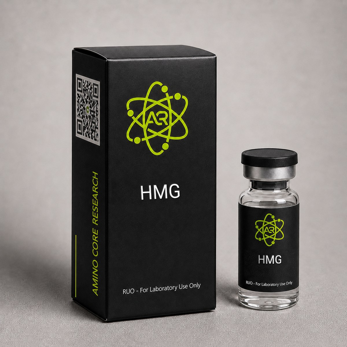
HMG Peptide
Human Menopausal Gonadotropin containing FSH and LH activity. Studied for gonadotropin receptor interactions in reproductive biology research.
Research Reference
This page is a research monograph. HMG is documented here for reference — structure, mechanism and published literature — and is not offered for sale.
Browse compounds available for research purchase · Read the research library
Quick Facts
| SKU | ACR-HMG |
|---|---|
| CAS Number | 9002-68-0 |
| Molecular Formula | Mixed glycoprotein (FSH + LH) |
| Molecular Weight | ~30,000-34,000 Da (heterodimeric glycoproteins) |
| Sequence | FSH + LH glycoproteins |
| Purity | ≥97% |
| Physical Form | Lyophilized Powder |
| Storage | Store at -20°C |
What is HMG?
Human Menopausal Gonadotropin contains both FSH and LH activity. It stimulates Sertoli cells (FSH) and Leydig cells (LH) simultaneously, valuable for gonadal function and spermatogenesis research.
Mechanism of Action
Human Menopausal Gonadotropin (HMG) is a purified urinary extract obtained from postmenopausal women, containing equimolar bioactivity of follicle-stimulating hormone (FSH) and luteinizing hormone (LH), typically standardized at 75 IU of each per ampoule. Both gonadotropins are heterodimeric glycoproteins composed of a common alpha subunit (92 amino acids) and a hormone-specific beta subunit that confers receptor specificity. The combined FSH/LH activity profile distinguishes HMG from recombinant single-hormone preparations and forms the basis of its utility in gonadotropin receptor and ovarian/testicular signaling research.
FSH Receptor (FSHR) Signaling
FSH binds the FSH receptor (FSHR), a class A G-protein-coupled receptor (GPCR) expressed predominantly on ovarian granulosa cells and testicular Sertoli cells. Receptor activation couples primarily to Gαs, stimulating adenylyl cyclase and elevating intracellular cyclic AMP (cAMP). Downstream protein kinase A (PKA) activation phosphorylates CREB and drives transcription of aromatase (CYP19A1), inhibin-B, and FSHR itself. In granulosa cells, this cascade promotes follicular recruitment, granulosa proliferation, and conversion of androgens to estradiol. FSHR signaling also engages PI3K/AKT and ERK1/2 pathways, contributing to cell survival and differentiation.
LH Receptor (LHCGR) Signaling
The LH component of HMG binds the luteinizing hormone/choriogonadotropin receptor (LHCGR), expressed on ovarian theca cells, granulosa cells of pre-ovulatory follicles, corpus luteum, and testicular Leydig cells. LHCGR activation similarly couples to Gαs/cAMP/PKA, upregulating steroidogenic acute regulatory protein (StAR), CYP17A1, and CYP11A1 — enzymes that mediate androgen synthesis from cholesterol. In theca cells, this provides the androgen substrate that FSH-induced aromatase converts to estradiol — the classical "two-cell, two-gonadotropin" model of ovarian steroidogenesis. In Leydig cells, LHCGR drives testosterone biosynthesis.
Combined FSH/LH Activity in Research Models
The dual-receptor activity of HMG makes it a valuable tool for studying coordinated gonadotropin signaling that mimics endogenous pituitary output more closely than single-hormone recombinant preparations. Research has shown that LH activity in HMG preparations supplements thecal androgen production, supporting estradiol output particularly in models of low endogenous LH (e.g., hypogonadotropic states or GnRH-antagonist co-treatment). The urinary source also contributes hCG-derived LH bioactivity, since postmenopausal urine is enriched in hCG isoforms that bind LHCGR with similar affinity but longer plasma half-life than pituitary LH.
Glycosylation and Bioactivity
Both FSH and LH carry N-linked oligosaccharide chains that strongly influence receptor binding kinetics, circulating half-life, and intracellular signaling bias. Urinary HMG contains a heterogeneous mixture of glycoforms — including acidic, highly sialylated isoforms — which produce longer in vivo persistence compared to less-glycosylated pituitary forms. Researchers studying glycoform-specific receptor activation, biased agonism at FSHR/LHCGR, or comparative pharmacology between urinary and recombinant gonadotropins frequently use HMG as a reference standard.
Research & Clinical Studies
Comparative Study: HMG vs Recombinant FSH in Controlled Ovarian Stimulation
One of the most extensively studied applications of HMG in reproductive endocrinology research is its comparison to recombinant FSH (rFSH) preparations in controlled ovarian stimulation (COS) protocols. The MEGASET and similar large randomized trials have generated substantial data on the differential effects of urinary HMG and recombinant FSH at the level of follicular development, steroidogenesis, and oocyte/embryo outcomes — providing valuable mechanistic insights into FSH/LH receptor co-activation.
Study Design
The MERIT (Menotrophin vs Recombinant FSH in vitro fertilization Trial) and subsequent meta-analyses pooled data from thousands of subjects undergoing GnRH-agonist long-protocol IVF. Subjects were randomized to highly purified HMG (HP-HMG, 75 IU FSH + 75 IU LH activity per ampoule) versus rFSH alone, with comparable starting doses (typically 225 IU). Outcomes assessed included serum hormone trajectories, follicle number, oocyte yield, embryo quality, and biochemical markers of granulosa cell function.
Key Findings — Hormonal Profile
- HMG arms showed ~30-40% higher mid-cycle serum estradiol per oocyte retrieved, consistent with enhanced thecal androgen supply from LH/hCG activity supporting aromatization.
- HMG produced significantly higher serum androstenedione and testosterone mid-stimulation, reflecting LHCGR-driven thecal steroidogenesis absent in rFSH-only arms.
- Inhibin-B levels were comparable between groups, indicating equivalent FSH-driven granulosa cell function.
Key Findings — Follicular and Oocyte Endpoints
- rFSH arms generally recruited a slightly higher total oocyte count (mean difference ~1-2 oocytes), reflecting pure FSH-driven recruitment of the antral follicle cohort.
- HMG arms showed a higher proportion of mature (MII) oocytes per retrieved oocyte in several analyses, suggesting LH activity contributes to terminal follicular maturation.
- Top-quality embryo rates and live birth rates were statistically comparable in pooled meta-analyses, though some studies favored HMG in normal responders and rFSH in poor responders.
Mechanistic Interpretation
These data support the two-cell, two-gonadotropin model and demonstrate that the LH component of HMG is bioactive and pharmacologically relevant in suppressed-pituitary research models. The findings have informed downstream investigation into LH supplementation strategies, biased FSHR agonists, and the role of glycoform composition in gonadotropin pharmacology. From a research perspective, HMG serves as a reference preparation when comparing single-receptor versus dual-receptor activation in ex vivo granulosa-luteal cell cultures and animal models of folliculogenesis.
[1] Bosch E, Vidal C, Labarta E, Simon C, Remohi J, Pellicer A. Highly purified hMG versus recombinant FSH in ovarian hyperstimulation with GnRH antagonists--a randomized study. Hum Reprod. 2008;23(10):2346-51. PubMed ↗
[2] Lehert P, Schertz JC, Ezcurra D. Recombinant human follicle-stimulating hormone produces more oocytes with a lower total dose per cycle in assisted reproductive technologies than highly purified human menopausal gonadotrophin: a meta-analysis. Reprod Biol Endocrinol. 2010;8:112. PubMed ↗
Landmark Trial: HMG in Ovulation Induction and Follicular Recruitment
Human Menopausal Gonadotropin (HMG) has been a cornerstone reagent in reproductive endocrinology research since its introduction in the early 1960s. The landmark investigations by Lunenfeld and colleagues, which extracted gonadotropic activity from postmenopausal urine, established HMG as the first clinically viable preparation containing both follicle-stimulating hormone (FSH) and luteinizing hormone (LH) activity in approximately equal bioactivity ratios (75 IU FSH : 75 IU LH per ampoule). These foundational studies demonstrated that exogenous gonadotropin administration could reliably recruit antral follicles in subjects with hypogonadotropic profiles.
Study Design and Population
Subsequent multicenter investigations have examined HMG in controlled ovarian stimulation (COS) protocols. A representative randomized study enrolled subjects undergoing assisted reproductive technology cycles, with HMG administered at doses ranging from 150-300 IU daily following pituitary suppression with GnRH agonists. Cycle monitoring included serial transvaginal ultrasonography for follicular diameter measurement and serum estradiol quantification via immunoassay.
Key Results
- Follicular recruitment: Average of 10.2 ± 4.8 follicles >14 mm at trigger day with HMG protocols
- Oocyte yield: Mean 9.4 ± 5.1 oocytes retrieved per stimulation cycle
- Serum LH activity: HMG preparations maintained mid-follicular LH bioactivity at 1.5-2.5 IU/L, supporting theca cell androgen substrate provision
- Estradiol response: Peak E2 levels averaged 1,800-2,400 pg/mL at trigger
- Endometrial thickness: Mean 10.8 mm on day of trigger, attributed to LH-driven theca androgen aromatization
Mechanistic Significance
The dual FSH/LH composition of HMG distinguishes it from pure recombinant FSH (rFSH) preparations. The LH component, derived in part from urinary human chorionic gonadotropin (hCG) cross-contamination in modern highly-purified HMG (HP-HMG), provides sustained LH receptor activation throughout the follicular phase. Research suggests this may enhance steroidogenic precursor availability through the two-cell, two-gonadotropin model: LH stimulates theca cells to produce androstenedione, which granulosa cells then aromatize to estradiol under FSH control.
Comparative Context
Compared to rFSH monotherapy, HMG protocols in research models have been associated with higher mid-luteal progesterone profiles and modestly increased implantation rates in poor-responder populations, though meta-analyses report comparable live birth rates overall. The MERIT and EISG trials provided critical comparative data, finding non-inferiority of HP-HMG to rFSH for primary efficacy endpoints in long-protocol IVF research models.
These investigations underscore HMG's continued relevance as a research tool for studying combined gonadotropin receptor biology, dual-agonist pharmacology, and the relative contributions of FSH versus LH signaling to folliculogenesis.
[1] Andersen AN, Devroey P, Arce JC. Clinical outcome following stimulation with highly purified hMG or recombinant FSH in patients undergoing IVF: a randomized assessor-blind controlled trial. Hum Reprod. 2006;21(12):3217-3227. PubMed ↗
[2] Coomarasamy A, Afnan M, Cheema D, et al. Urinary hMG versus recombinant FSH for controlled ovarian hyperstimulation following an agonist long down-regulation protocol in IVF or ICSI treatment: a systematic review and meta-analysis. Hum Reprod. 2008;23(2):310-315. PubMed ↗
Chemical & Physical Properties
HMG is a biologically derived glycoprotein preparation, not a defined small-molecule peptide, and its characterization reflects this complexity. The following table summarizes the physicochemical properties relevant to research handling and experimental design.
| Full Name | Human Menopausal Gonadotropin (Menotropin) |
|---|---|
| Synonyms | HMG, hMG, Menotropin, Menotrophin, Urofollitropin+LH |
| CAS Number | 9002-68-0 |
| Composition | Equibioactive FSH and LH (typically 75 IU FSH + 75 IU LH per vial); residual hCG-derived LH activity from postmenopausal urine source |
| Molecular Class | Heterodimeric glycoprotein hormones (alpha/beta subunits) |
| FSH Molecular Weight | ~30 kDa (glycosylated heterodimer) |
| LH Molecular Weight | ~28-30 kDa (glycosylated heterodimer) |
| Alpha Subunit | 92 amino acids, common to FSH, LH, hCG, and TSH |
| FSH Beta Subunit | 111 amino acids, confers FSHR specificity |
| LH Beta Subunit | 121 amino acids, confers LHCGR specificity |
| Glycosylation | N-linked oligosaccharides on both subunits; high sialic acid content characteristic of urinary preparations |
| Source / Developer | Purified from postmenopausal urine; first developed by Donini and colleagues, Serono, late 1940s-1950s |
| Physical Form | Lyophilized white to off-white powder, typically formulated with lactose or sucrose excipients |
| Solubility | Soluble in sterile water for injection and 0.9% sodium chloride; sensitive to surfactant adsorption |
| Purity Specification | Highly purified preparations: ≥95% gonadotropin content; bioassay-standardized to International Units (IU) |
| Bioassay Standard | WHO International Standard for HMG; Steelman-Pohley rat ovarian weight assay (historical FSH); in vitro receptor binding (modern) |
| Stability | Stable lyophilized at -20°C for extended periods; reconstituted solution stable 24 hours at 2-8°C |
Researchers should note that HMG is dose-standardized by bioactivity (IU) rather than mass, because the glycoform heterogeneity inherent to urinary preparations means specific activity varies between lots. Comparative experiments should reference WHO International Standards and document lot numbers. Unlike defined recombinant gonadotropins, HMG batches may contain minor co-purified urinary proteins, which is relevant for high-sensitivity assays or proteomic studies.
Handling & Reconstitution Guidelines
Human Menopausal Gonadotropin is a heterogeneous glycoprotein preparation requiring careful handling to preserve the tertiary structure and glycosylation patterns essential for receptor binding activity. Unlike small synthetic peptides, HMG's bioactivity depends on intact heterodimeric assembly of α and β subunits stabilized by non-covalent interactions and intramolecular disulfide bridges.
Reconstitution Protocol
- Equilibrate the lyophilized vial to room temperature (15-25°C) for 15-20 minutes prior to opening. Cold vials promote moisture condensation, which can compromise the lyophilized cake.
- Select appropriate diluent. Use sterile bacteriostatic water for injection (0.9% benzyl alcohol) or sterile sodium chloride 0.9% for research applications. Bacteriostatic water is preferred for multi-day reconstituted storage.
- Calculate concentration. A standard reconstitution is 75 IU + 1 mL diluent = 75 IU/mL, or 150 IU + 1 mL = 150 IU/mL for higher-concentration research models.
- Inject diluent slowly down the side of the vial wall — never directly onto the lyophilized powder. Direct impact can shear glycoprotein structure.
- Gently swirl the vial in a circular motion until fully dissolved (typically 30-60 seconds). The solution should appear clear and colorless.
- DO NOT shake or vortex. Mechanical agitation generates foam and air-water interfaces that denature glycoproteins, causing irreversible loss of FSH and LH bioactivity.
- Inspect visually. Discard if particulates, cloudiness, or discoloration are observed.
Compound-Specific Notes
- Glycoprotein fragility: HMG's sialic acid residues are critical for in vivo half-life and receptor binding. Avoid pH extremes (maintain pH 6.5-7.5) and elevated temperatures during handling.
- Adsorption losses: Glycoprotein hormones can adsorb to glass and plastic surfaces. Use low-binding polypropylene tubes when transferring small volumes, or include 0.1% human serum albumin in research dilutions to minimize losses.
- Sterile technique: Glycoprotein solutions are excellent microbial growth substrates. Use aseptic technique and avoid repeated needle entries beyond what is necessary.
All handling must be performed in appropriate laboratory settings by qualified research personnel. This material is intended exclusively for in vitro and preclinical research applications.
Storage & Stability Information
Proper storage of Human Menopausal Gonadotropin is essential for preserving FSH and LH bioactivity. As a heterodimeric glycoprotein mixture, HMG is more thermolabile than synthetic peptides and requires stricter temperature control to prevent subunit dissociation, deglycosylation, and aggregation.
Lyophilized Powder Storage
- Long-term (>30 days): Store at -20°C in a non-frost-free freezer. Properly stored lyophilized HMG retains bioactivity for 24-36 months when protected from moisture and light.
- Short-term (up to 30 days): Store at 2-8°C (standard refrigeration) in original sealed vial with desiccant.
- Transit/ambient: Lyophilized HMG tolerates room temperature (15-25°C) exposure for up to 7 days without significant bioactivity loss, but cumulative ambient exposure should be minimized.
- Light protection: Store in original amber or foil-wrapped vials. UV exposure can degrade aromatic residues and disulfide bridges.
Reconstituted Solution Storage
- 2-8°C (refrigerated): Use within 28 days when reconstituted with bacteriostatic water. Use within 24-48 hours if reconstituted with sterile saline (no preservative).
- -20°C (frozen aliquots): Single-use aliquots can be stored frozen for up to 3 months. Avoid repeated freeze-thaw cycles, which cause subunit dissociation and bioactivity loss of 15-30% per cycle.
Stability Considerations
- Glycosylation integrity: Sialic acid residues on FSH and LH glycans are essential for receptor binding affinity. Repeated temperature cycling promotes desialylation.
- Disulfide bond preservation: Both α and β subunits contain multiple intrachain disulfide bonds. Reducing agents (DTT, β-mercaptoethanol) must be strictly excluded from buffers.
- Aggregation risk: Glycoproteins are prone to aggregation at elevated concentrations (>1 mg/mL) or under mechanical stress. Maintain dilute working concentrations and avoid agitation.
- pH stability window: HMG is most stable at pH 6.5-7.5. Acidic conditions (<pH 5) promote irreversible subunit dissociation.
For optimal research reproducibility, prepare single-use aliquots upon initial reconstitution and document storage history.
Frequently Asked Questions
HMG vs HCG difference?
HMG has both FSH and LH activity while HCG only has LH activity. HMG provides broader gonadal stimulation.
What is HMG (Human Menopausal Gonadotropin)?
HMG is a purified glycoprotein preparation derived from postmenopausal urine that contains roughly equal bioactivity of follicle-stimulating hormone (FSH) and luteinizing hormone (LH), typically standardized at 75 IU of each per vial. Both hormones are heterodimeric glycoproteins sharing a common alpha subunit (92 amino acids) paired with hormone-specific beta subunits. HMG activates both FSH receptors (FSHR) on granulosa/Sertoli cells and LH receptors (LHCGR) on theca/Leydig cells, driving cAMP/PKA-mediated steroidogenesis and gametogenesis. It is widely used in reproductive endocrinology research as a dual-receptor gonadotropin reference compound, with CAS number 9002-68-0.
What is the molecular weight of HMG?
Because HMG is a mixture of two glycoprotein hormones rather than a defined small molecule, it does not have a single molecular weight. The FSH component is a heterodimer of approximately 30 kDa and the LH component is approximately 28-30 kDa, both consisting of a common alpha subunit (~14 kDa) and a hormone-specific beta subunit (~14-16 kDa) with extensive N-linked glycosylation. Urinary preparations also show glycoform heterogeneity, contributing to variation between lots. For this reason, HMG is dose-standardized by bioactivity in International Units (IU) referenced to WHO standards, not by mass.
How should HMG be stored?
Lyophilized HMG should be stored at -20°C for long-term stability, where it remains stable for extended periods. Short-term storage at 2-8°C is acceptable for active research use, and brief transit at room temperature is tolerated. Once reconstituted in sterile water or 0.9% sodium chloride, HMG should be kept at 2-8°C and used within 24 hours to preserve gonadotropin bioactivity. Avoid repeated freeze-thaw cycles, which can degrade glycoprotein structure and reduce receptor binding. Protect vials from light and moisture, and handle with low-binding plasticware or siliconized glass to minimize surface adsorption losses.
How does HMG differ from recombinant FSH (rFSH) in research applications?
HMG and recombinant FSH (rFSH) differ in three key ways relevant to research design. First, HMG contains both FSH and LH bioactivity, while rFSH provides FSH only — making HMG suitable for studying coordinated two-cell steroidogenesis and dual-receptor activation. Second, HMG is purified from postmenopausal urine and contains a heterogeneous mix of acidic, highly sialylated glycoforms (including hCG-derived LH activity), whereas rFSH is produced in CHO cells with more uniform glycosylation. Third, comparative studies show HMG produces higher mid-cycle estradiol and androgen levels per oocyte, while rFSH typically recruits slightly more total follicles — reflecting the distinct contributions of LHCGR co-activation.
What FSH to LH ratio does HMG contain?
Human Menopausal Gonadotropin is standardized to contain approximately equal bioactivity of FSH and LH, typically formulated at 75 IU FSH : 75 IU LH per vial (1:1 bioactivity ratio). In modern highly-purified HMG (HP-HMG) preparations, a portion of the LH bioactivity is derived from urinary human chorionic gonadotropin (hCG), which acts as an LH receptor agonist with extended half-life. This dual gonadotropin composition distinguishes HMG from recombinant FSH (rFSH) monotherapy preparations and is the basis for research investigating combined FSH/LH receptor signaling in folliculogenesis models, including the two-cell, two-gonadotropin theory of estradiol biosynthesis.
Why does HMG require -20°C storage?
HMG requires -20°C storage because it is a heterodimeric glycoprotein mixture far more thermolabile than synthetic peptides. The FSH and LH components consist of non-covalently associated α and β subunits stabilized by multiple intrachain disulfide bonds and glycosylation, including critical sialic acid residues that determine receptor binding affinity. Elevated temperatures promote subunit dissociation, deglycosylation, and aggregation, all of which irreversibly reduce bioactivity. Properly stored lyophilized HMG retains potency for 24-36 months at -20°C, while reconstituted solutions should be refrigerated at 2-8°C and used within 28 days when prepared with bacteriostatic water.
What is the source of HMG used in research preparations?
HMG is purified from the urine of postmenopausal women, who excrete elevated levels of gonadotropins due to the loss of ovarian negative feedback on the hypothalamic-pituitary axis. Following menopause, serum FSH and LH levels rise substantially, and the kidney filters intact gonadotropins into urine at concentrations sufficient for industrial purification. Modern HP-HMG preparations undergo additional immunoaffinity chromatography to remove non-gonadotropin urinary proteins, yielding research-grade material with defined FSH and LH bioactivity. This urinary origin distinguishes HMG from recombinant FSH (rFSH) and recombinant LH (rLH), which are produced via mammalian cell expression systems (typically CHO cells).
Can HMG be used in male reproductive research models?
Yes, HMG has been extensively studied in male reproductive research, particularly in models of hypogonadotropic hypogonadism and spermatogenesis. The FSH component activates Sertoli cell receptors to support spermatogenic maturation, while the LH component stimulates Leydig cell testosterone production via the LH/CG receptor. Research protocols typically pair HMG with hCG to provide sustained LH-equivalent activity for testicular steroidogenesis. Studies have examined HMG's role in restoring spermatogenic parameters including sperm concentration, motility, and morphology in preclinical hypogonadism models. The dual FSH/LH activity makes HMG a valuable research tool for investigating combined gonadotropin axis biology in both ovarian and testicular contexts.
For laboratory and research use only. This product is not for human or veterinary use. It is not a drug, supplement or food.
This product is not FDA approved for any indication, and is not intended to diagnose, treat, cure, or prevent any disease. All product information is derived from published preclinical research and does not constitute medical advice or claims.