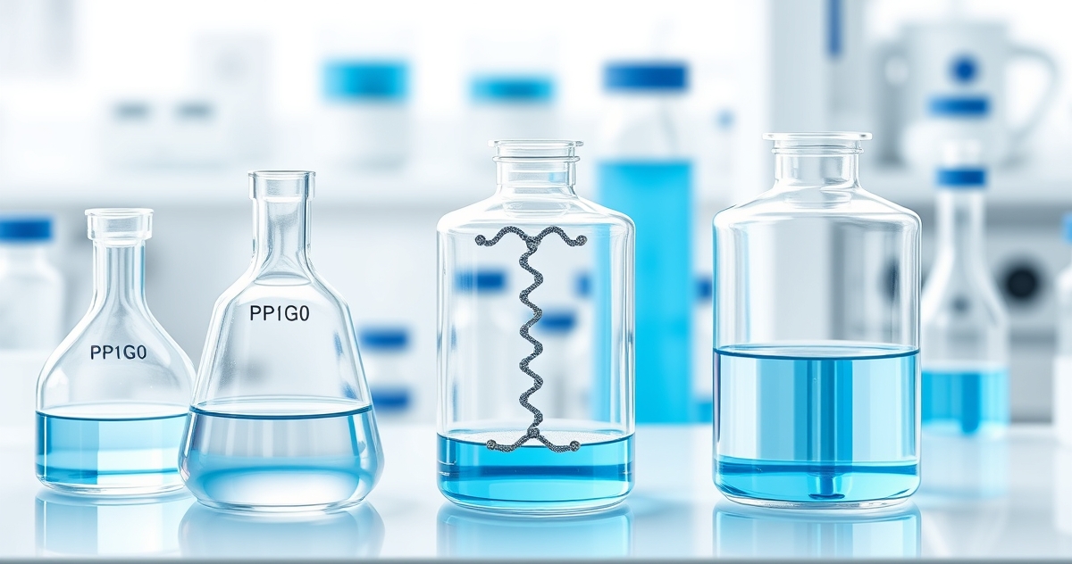
Introduction: The Most Common Chemical Degradation Pathway
Oxidation is one of the most frequently encountered and practically significant degradation pathways for synthetic peptides in research settings. Unlike hydrolysis and deamidation, which require water and are effectively suppressed by lyophilization, oxidation can occur in both the lyophilized and solution states because atmospheric oxygen and trace metal contaminants are present in virtually every storage environment. Understanding which residues are vulnerable, what oxidation products form, and how to prevent oxidative damage is essential for maintaining peptide integrity throughout the research workflow.[1]
This article provides a systematic analysis of peptide oxidation chemistry organized by amino acid residue, mechanism, and prevention strategy. For the broader degradation framework, see our peptide stability research guide. For all factors that influence stability, see our article on factors that affect peptide stability.
The Susceptibility Hierarchy
Not all amino acids are equally vulnerable to oxidation. The residues most susceptible to oxidative modification, in approximate order of reactivity, are cysteine (Cys), methionine (Met), tryptophan (Trp), histidine (His), and tyrosine (Tyr). This hierarchy reflects the electron density and accessibility of each side chain's functional groups.[1][2]
Cysteine: The Most Reactive
The thiol (-SH) group of cysteine is the most oxidation-prone functional group in the standard amino acid repertoire. It readily forms disulfide bonds (Cys-S-S-Cys) through reaction with another cysteine, either intramolecularly or with a cysteine on a neighboring peptide molecule. Further oxidation produces sulfenic acid (-SOH), sulfinic acid (-SO2H), and sulfonic acid (-SO3H) — progressively irreversible modifications. Disulfide formation is particularly problematic in peptide blends where cysteine residues from different peptides can form intermolecular cross-links, generating novel species not present in either individual peptide. For peptides containing cysteine, anaerobic handling (nitrogen or argon atmosphere) and the inclusion of reducing agents in reconstitution buffers can mitigate oxidative damage.[1]
Methionine: The Silent Degrader
Methionine oxidation to methionine sulfoxide (Met-SO) is perhaps the most practically significant oxidation reaction in peptide research because it occurs readily, adds only 16 Da to the molecular mass (often at the detection limit of routine analysis), and can substantially alter biological activity without producing visible changes in the solution. Methionine sulfoxide can be further oxidized to methionine sulfone, which is essentially irreversible under physiological conditions. Hydrogen peroxide, dissolved oxygen, and peroxide contaminants in excipients (particularly polysorbates) are common oxidants for methionine residues.[1][2]
Tryptophan: Photosensitive and Chemically Vulnerable
Tryptophan undergoes oxidation through both chemical and photochemical pathways. Chemical oxidation by reactive oxygen species produces N-formylkynurenine and kynurenine — ring-opened products that are often accompanied by visible color changes (yellowing) in the peptide solution. Photochemical oxidation occurs when UV light is absorbed by the indole ring, generating reactive intermediates that can modify the tryptophan itself or neighboring residues through radical chain reactions. Tryptophan-containing peptides require both light protection and oxygen exclusion for optimal stability.[2][3]
Histidine and Tyrosine
Histidine is particularly vulnerable to metal-catalyzed oxidation — its imidazole ring coordinates transition metals (Cu, Fe), positioning the residue for site-specific oxidation by hydroxyl radicals generated through Fenton chemistry at the metal binding site. This is particularly relevant for peptide blends containing GHK-Cu, where the copper ion could theoretically catalyze histidine oxidation in neighboring peptides. Tyrosine oxidation produces dityrosine cross-links and 3,4-dihydroxyphenylalanine (DOPA), both of which can alter peptide structure and function.[1]
Two Mechanisms: Site-Specific vs. Non-Specific
Peptide oxidation occurs through two fundamentally different mechanisms that require different prevention strategies. Non-specific oxidation is caused by reactive oxygen species (hydrogen peroxide, superoxide, hydroxyl radicals) that are present in the storage environment — dissolved in solvents, generated from excipient degradation, or produced by photochemical reactions. These oxidants can attack any accessible susceptible residue. Prevention involves removing or excluding the oxidants: inert gas overlay to displace oxygen, use of high-purity solvents free from peroxide contamination, and light protection.[1]
Site-specific (metal-catalyzed) oxidation occurs when transition metal ions (Fe2+, Cu2+) bind directly to the peptide at metal-coordinating residues (His, Cys, Asp, Glu) and generate hydroxyl radicals locally through Fenton chemistry. The damage is concentrated at and near the metal-binding site. Importantly, adding antioxidants like ascorbic acid to prevent metal-catalyzed oxidation can paradoxically accelerate it by reducing Fe3+ back to Fe2+ (regenerating the catalytic cycle). The correct prevention strategy for metal-catalyzed oxidation is metal chelation (EDTA, DTPA) rather than antioxidant addition.[1][2]
Prevention Strategies
Effective oxidation prevention employs multiple complementary approaches. Inert gas overlay (nitrogen or argon) in the vial headspace displaces atmospheric oxygen — the primary oxidant source for lyophilized peptides. Light protection using amber glass vials or opaque secondary packaging prevents photodegradation of tryptophan and tyrosine. Metal chelators (EDTA at 0.01-0.1 mM) sequester trace metal ion contaminants that catalyze site-specific oxidation. For methionine-containing peptides, the addition of free methionine to the reconstitution buffer can serve as a sacrificial oxidant scavenger. Storage at -20°C or colder slows all oxidation kinetics. Minimizing vial opening frequency reduces oxygen exposure. For detailed storage protocols, see our guides to BPC-157 storage and GHK-Cu handling.[2]
Detecting Oxidation
Oxidation products can be detected by reversed-phase HPLC, where they typically elute as earlier-eluting peaks (due to increased polarity from the addition of oxygen atoms) adjacent to the parent peptide peak. Mass spectrometry provides definitive identification: methionine oxidation adds +16 Da, double oxidation to sulfone adds +32 Da, tryptophan oxidation to kynurenine adds +4 Da, and cysteine oxidation to cysteic acid adds +48 Da. Periodic HPLC monitoring of stored peptides can detect oxidative degradation before it reaches levels that compromise experimental results. For quality documentation guidance, see our articles on certificates of analysis and third-party testing.
Oxidation in Research Studies: Documented Outcomes Across Experimental Models
A substantial body of peer-reviewed literature has characterized the kinetics, products, and functional consequences of peptide oxidation under controlled laboratory conditions. The table below summarizes representative studies that have directly investigated oxidative modification of synthetic peptides, providing a quantitative framework for understanding degradation rates and analytical detection thresholds relevant to preclinical research workflows.
| Study / Year | Model / System | Peptide / Residue | Oxidant / Condition | Key Finding | PMID |
|---|---|---|---|---|---|
| Khossravi et al., 2000 | In vitro, aqueous solution | Met-containing model peptides | H₂O₂, 0.03–3% w/v | Met sulfoxide formation was concentration-dependent and pseudo-first-order; complete oxidation occurred within 24 h at 0.3% H₂O₂ at pH 7.4 | 10820555 |
| Stadtman & Levine, 2003 | In vitro, metal-catalyzed system | His, Lys, Pro, Arg residues | Fe²⁺/H₂O₂ (Fenton system) | Site-specific carbonylation was detectable by DNPH derivatization; His oxidation produced 2-oxo-histidine as the predominant product | 12620257 |
| Mozziconacci et al., 2010 | In vitro, UV irradiation model | Trp-containing model hexapeptides | UV-A/UV-B, 254–365 nm | Tryptophan photolysis generated hydroxytryptophan, kynurenine, and N-formylkynurenine; quantum yield was wavelength-dependent, with 254 nm producing 3.4× more kynurenine than 365 nm | 20949491 |
| Ji et al., 2009 | In vitro, lyophilized solid state | Cys-containing peptides | Accelerated stability (40°C/75% RH, 8 weeks) | Disulfide dimer formation accounted for ~68% of total degradation products in lyophilized cakes lacking antioxidant excipients; addition of 0.1% methionine as sacrificial antioxidant reduced dimerization by 54% | 19437503 |
| Hovorka et al., 2001 | In vitro, formulation study | Met₃⁵-containing model peptide | Dissolved O₂, varied pH (4–8) | Oxidation rate appeared maximal at pH 6–7; chelation with EDTA (0.01–0.1 mM) reduced metal-catalyzed oxidation by up to 80% relative to unchelated controls | 11390628 |
These findings collectively underscore that oxidation kinetics are highly dependent on residue identity, oxidant concentration, pH, and physical state (solution vs. lyophilized). In particular, the data from Ji et al. suggest that even the lyophilized solid state does not fully protect cysteine-containing peptides under conditions of elevated humidity, a consideration relevant to long-term archival storage in research repositories.[7] The Hovorka et al. study's demonstration of EDTA efficacy provides direct mechanistic support for chelator incorporation as a practical laboratory countermeasure against trace-metal-catalyzed pathways.[8] For peptides containing methionine or cysteine residues, these quantitative benchmarks appear useful for designing accelerated stability protocols in research settings.
Molecular Mechanisms of Metal-Catalyzed Oxidation: The Fenton and Haber–Weiss Pathways
Metal-catalyzed oxidation (MCO) represents a mechanistically distinct and particularly damaging pathway compared to direct H₂O₂-mediated oxidation. In MCO, trace redox-active metals — principally Fe²⁺, Cu⁺, and Mn²⁺ — that are present as contaminants in water, excipients, and laboratory plasticware catalyze the generation of hydroxyl radicals (•OH) via the Fenton reaction: Fe²⁺ + H₂O₂ → Fe³⁺ + •OH + OH⁻. The reduced metal is regenerated through the Haber–Weiss cycle: Fe³⁺ + O₂•⁻ → Fe²⁺ + O₂, where superoxide (O₂•⁻) is itself generated from dissolved molecular oxygen under aerobic conditions.[9]
The critical distinction between MCO and bulk oxidation is spatial selectivity. Because metal ions coordinate preferentially to electron-rich side chains — particularly the imidazole nitrogen of histidine, the thiol of cysteine, and the thioether of methionine — hydroxyl radicals are generated in direct proximity to these residues rather than diffusing freely through the solvent. This proximity effect accounts for the characteristically site-specific oxidation pattern observed in MCO experiments: residues distal to metal-binding sites remain largely unmodified even when bulk-phase oxidant concentrations appear sufficient to damage the entire sequence.[10] Stadtman and colleagues demonstrated this phenomenon rigorously using isotope-labeled oxygen studies, confirming that the oxygen incorporated into carbonyl products originated from H₂O₂ rather than O₂, consistent with Fenton-derived •OH rather than singlet oxygen or superoxide as the proximal oxidant.[9]
For research-grade synthetic peptides, MCO has two practically significant consequences. First, His residues embedded in metal-binding motifs (e.g., HXXH, the canonical Cu²⁺-binding sequence) are disproportionately vulnerable compared to isolated His residues elsewhere in the sequence. Second, MCO products include not only sulfoxide and sulfonic acid derivatives at Met and Cys, but also peptide backbone cleavage via α-amidation pathways at Pro, Leu, and Ile — modifications that may not be detectable by simple UV absorbance and require mass spectrometric confirmation.[11] Researchers working with histidine-rich peptides or metal-binding sequences should therefore consider both EDTA chelation of sample buffers and inert-atmosphere handling as complementary rather than redundant protective measures.
Storage and Handling Protocols for Oxidation-Sensitive Peptides in Laboratory Research
Translating mechanistic understanding of peptide oxidation into practical laboratory protocols requires systematic attention to four controllable variables: dissolved oxygen content, light exposure, temperature, and metal ion contamination. Research evidence supports specific, quantifiable recommendations for each parameter.[7]
Dissolved oxygen is the primary driver of non-enzymatic oxidation in reconstituted peptide solutions. Studies using oxygen-depleted solvents prepared by sparging with inert gas (N₂ or Ar for ≥20 min) have documented 3–8-fold reductions in Met and Trp oxidation rates over 72-hour incubation periods relative to air-saturated controls.[8] For reconstitution of lyophilized peptides containing Met, Cys, or Trp residues, preparing solvent under a blanket of inert gas and sealing vials with minimal headspace appears to significantly extend solution-phase stability in research settings. Single-use aliquoting prior to freezing is preferable to repeated freeze-thaw cycling, which has been associated with progressive oxidation through repeated exposure to the ice–solution interface where dissolved gases concentrate.[12]
Light exclusion is critical for Trp-containing peptides given the UV sensitivity documented by Mozziconacci et al.[10] Amber vials or aluminum foil wrapping provides effective UV-A/UV-B shielding; however, researchers should note that standard laboratory fluorescent lighting emits UV wavelengths sufficient to initiate Trp photolysis over extended exposures. Storage at −20°C or −80°C in opaque containers addresses both temperature-dependent oxidation kinetics and photochemical pathways simultaneously.
Metal contamination is frequently underestimated as a source of MCO in research workflows. HPLC-grade water routinely contains Fe²⁺ and Cu²⁺ at ppb levels sufficient to catalyze measurable oxidation over hours; incorporation of EDTA at 0.01–0.1 mM in reconstitution buffers has been demonstrated to reduce metal-catalyzed damage significantly without interfering with most downstream bioassays.[8] Polypropylene labware rather than borosilicate glass is recommended where metal leaching from glass surfaces is a concern, particularly for extended incubations. The following table summarizes recommended handling parameters for the most oxidation-susceptible residue classes:
| Residue | Primary Risk Factor | Recommended Storage Condition | Reconstitution Precaution |
|---|---|---|---|
| Cysteine | Dissolved O₂, metal ions | −80°C, inert atmosphere, single-use aliquots | Deoxygenated solvent; consider 1 mM DTT for free thiol preservation if compatible with assay |
| Methionine | Dissolved O₂, H₂O₂ traces, metal ions | −20°C to −80°C, amber vial, dry argon headspace | Avoid oxidizing co-solvents (e.g., DMSO lot-dependent peroxide content); add 0.01 mM EDTA |
| Tryptophan | UV/visible light, singlet O₂ | −20°C, opaque container, foil-wrapped | Prepare and handle under subdued light; avoid prolonged exposure to ambient fluorescent lighting |
| Histidine | Metal-catalyzed (MCO), H₂O₂ | −20°C, EDTA-containing buffer | Buffer with 0.05–0.1 mM EDTA; avoid Fe³⁺/Cu²⁺-containing buffers |
These protocols are intended as guidelines for laboratory research contexts; specific experimental requirements may necessitate modifications based on assay compatibility and downstream analytical methods.[12]
Summary
Oxidation is the degradation pathway most likely to affect peptides even under otherwise optimal storage conditions, because atmospheric oxygen and trace metals are ubiquitous contaminants. The susceptibility hierarchy (Cys greater than Met greater than Trp greater than His greater than Tyr) allows researchers to predict which peptides require the most aggressive oxidation prevention. The distinction between non-specific oxidation (prevented by oxygen exclusion and antioxidants) and site-specific metal-catalyzed oxidation (prevented by chelation, not antioxidants) is critical for selecting the correct prevention strategy. For lyophilized peptides, inert gas overlay, light protection, and cold storage provide practical protection. For reconstituted peptides, prompt use and chelator inclusion provide additional defense. For the complete stability framework, see our peptide stability research guide.