Introduction: Recognizing Compromised Material
Peptide degradation is not always obvious. While some forms of degradation produce visible changes — color shifts, cloudiness, or altered powder appearance — many of the most consequential degradation reactions generate products that are visually indistinguishable from the intact peptide. A vial that looks perfectly clear and colorless may contain a mixture of deamidated, isomerized, and oxidized species with substantially reduced or altered biological activity. This invisible degradation is what makes quality awareness so important: researchers who rely solely on visual inspection may unknowingly use compromised material, generating irreproducible or misleading results.[1]
This article provides a comprehensive guide to recognizing degradation across three levels of assessment: what you can see, what instruments can detect, and what experimental behavior suggests. For the underlying degradation chemistry, see our peptide stability research guide. For understanding what causes degradation, see our article on factors that affect peptide stability.
Visual Indicators: What You Can See
Lyophilized (Powder) Form
A properly lyophilized peptide appears as a white to off-white, fluffy or loosely structured powder (the "cake") that occupies a defined volume in the vial. Signs that degradation or improper handling has occurred include cake collapse — the fluffy structure has condensed into a dense, glassy, or crystalline layer at the bottom of the vial, indicating that the glass transition temperature was exceeded (often due to moisture ingress or temperature excursion). Discoloration from white to yellow or brown suggests oxidation, particularly of tryptophan residues. A sticky, gummy, or wet appearance indicates moisture absorption, which accelerates all degradation pathways. Powder adhering to the stopper or cap rather than remaining at the bottom of the vial may indicate electrostatic issues or partial deliquescence.[1][2]
Not all visual changes indicate catastrophic degradation — some peptides are naturally off-white or slightly yellow, and minor variations in cake appearance between batches can reflect differences in lyophilization conditions rather than degradation. However, significant changes from the expected appearance, particularly darkening or liquefaction, warrant caution.
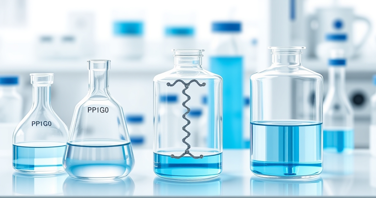
Reconstituted (Solution) Form
A properly reconstituted peptide should produce a clear, colorless (or very faintly tinted for copper-containing peptides like GHK-Cu) solution that is free of visible particles. Signs of degradation in reconstituted solution include cloudiness or turbidity, indicating aggregation or the formation of insoluble degradation products. Visible particulates — flocculent material, fibers, or granules that do not dissolve with gentle mixing — suggest advanced aggregation or precipitation. Color change to yellow or brown suggests oxidation of aromatic residues (tryptophan, tyrosine). Foaming that persists abnormally long after mixing may indicate surface-active degradation products. An unusual or foul odor suggests microbial contamination, which introduces proteases that rapidly degrade the peptide.[1]
Analytical Indicators: What Instruments Detect
HPLC Signatures
Reversed-phase HPLC is the most informative routine analytical method for detecting peptide degradation. Specific chromatographic indicators include a reduction in the main peak area compared with the original certificate of analysis, indicating loss of intact peptide. New peaks appearing earlier than the main peak (more hydrophilic) typically indicate oxidation products (methionine sulfoxide adds a polar oxygen atom) or deamidation products (conversion of neutral asparagine to charged aspartate). New peaks appearing later than the main peak (more hydrophobic) may indicate aggregation products or certain chemical modifications. Peak broadening or shouldering of the main peak suggests a mixture of closely related species — often the early stages of deamidation where the parent peptide co-elutes with its isoaspartate product.[3]
A purity decline of more than 2-3% from the original CoA value is generally considered significant and warrants reassessment of whether the peptide is suitable for continued use. A decline of more than 5-10% indicates substantial degradation that is likely to affect experimental results.
Mass Spectrometry Signatures
Mass spectrometry provides definitive identification of specific degradation products through characteristic mass shifts. Methionine oxidation produces a +16 Da shift. Deamidation of asparagine produces a +1 Da shift (detectable with high-resolution MS). Tryptophan oxidation to kynurenine produces a +4 Da shift. Cysteine oxidation to cysteic acid produces a +48 Da shift. Disulfide bond formation produces a -2 Da shift. Pyroglutamate formation from N-terminal glutamine produces a -17 Da shift. Chain cleavage produces fragments with masses corresponding to partial sequences.[3]
The presence of any of these mass-shifted species in a significant proportion (greater than 5% of total signal) indicates degradation that may affect research outcomes. For critical applications, third-party testing with both HPLC and MS provides the most comprehensive quality assessment.
Functional Indicators: What Experiments Reveal
Sometimes the first indication that a peptide has degraded comes not from inspection or analysis but from unexpected experimental results. Functional indicators of possible peptide degradation include a shift in the dose-response curve requiring higher concentrations to achieve previously observed effects — suggesting reduced potency of the intact peptide. Inconsistent results between experiments using different vials or aliquots of the same peptide — suggesting variable degradation across the stock. Loss of expected biological activity entirely — suggesting advanced degradation or the wrong peptide. Unexpected off-target effects not previously observed — suggesting that degradation products have different biological activity than the parent peptide. Failure to reproduce published results despite following the same protocol — when other variables have been controlled, peptide quality should be investigated.[1]
Including positive controls from freshly reconstituted, verified-quality peptide in every experiment provides the most reliable internal reference for detecting gradual potency loss over time.
Decision Framework: Use, Test, or Discard?
When degradation is suspected, researchers face a practical decision: continue using the material, invest in analytical testing, or discard and obtain fresh peptide. If visual degradation indicators are clearly present (cloudiness, discoloration, particulates), discard the material — the cost of fresh peptide is negligible compared with the cost of unreliable data. If the peptide looks normal but has been stored beyond recommended timelines, analytical testing by HPLC is recommended before use in critical experiments. If HPLC shows less than 2-3% decline from the original CoA, the peptide is generally acceptable for continued use. If HPLC shows 3-10% decline, the peptide may be acceptable for non-quantitative screening experiments but not for dose-response studies or other quantitative work. If HPLC shows greater than 10% decline, the peptide should be discarded regardless of visual appearance.
For peptides stored under recommended conditions (lyophilized at -20°C, sealed, dry), significant degradation within 12 months is unlikely for sequences without inherently labile residues. Beyond 12 months, the probability of measurable degradation increases and periodic re-testing becomes advisable. For all factors affecting stability and expected shelf-life timelines, see our dedicated articles.
Molecular Mechanisms of Peptide Degradation: Dominant Pathways and Sequence-Dependent Vulnerabilities
Understanding the chemical basis of peptide degradation allows researchers to anticipate which structural features render a given compound most susceptible to quality loss. Degradation does not proceed randomly; it is governed by specific reaction mechanisms that preferentially target defined amino acid residues, sequence motifs, and backbone configurations under predictable physicochemical conditions.
The four dominant degradation pathways in research peptide preparations are deamidation, oxidation, hydrolysis, and β-elimination/isomerization. Deamidation — the conversion of asparagine (Asn) or glutamine (Gln) to aspartate or glutamate via a succinimide intermediate — is arguably the most insidious pathway because it introduces only a +1 Da mass shift, which is below the resolution threshold of low-accuracy mass spectrometry instruments and produces no visual change whatsoever.[6] Sequences containing Asn-Gly or Asn-Ser dipeptide motifs are particularly vulnerable; the small glycine residue reduces steric hindrance around the succinimide transition state, accelerating deamidation rates by an order of magnitude relative to other Asn-X sequences.[7]
Oxidation disproportionately affects methionine (Met), tryptophan (Trp), cysteine (Cys), and histidine (His) residues. Methionine sulfoxidation (+16 Da) proceeds readily in the presence of dissolved oxygen, trace metal contaminants, or reactive oxygen species generated by UV photolysis. A study by Chou et al. (2005) demonstrated that methionine residues exposed on the surface of model peptides reached >40% oxidation within 48 hours when stored in aqueous solution at ambient temperature in the absence of antioxidant excipients or inert-atmosphere headspace.[8] For researchers working with Met-containing peptides such as those in the melanocortin or growth hormone-releasing families, this has direct implications for reconstitution protocols and storage vessel selection.
Backbone hydrolysis at Asp-Pro bonds deserves particular mention. This sequence motif is uniquely susceptible to acid-catalyzed hydrolysis due to the restricted rotation imposed by the proline pyrrolidine ring, which destabilizes the adjacent amide bond. Under mildly acidic reconstitution conditions (pH 3.0–4.5), Asp-Pro cleavage can generate truncated fragments within days — fragments that may retain partial receptor affinity and confound dose-response experiments.[6] Researchers using peptides containing this motif should monitor their material by HPLC with particular attention to new peaks in the 0.1–0.5 kDa retention window consistent with short N-terminal fragments.
Finally, disulfide scrambling in cysteine-containing peptides represents a structurally catastrophic yet analytically subtle form of degradation. Under alkaline reconstitution conditions or in the presence of trace thiols, native disulfide bonds can undergo thiol-disulfide exchange, producing non-native isomers with identical molecular masses but markedly different three-dimensional structures and biological activity profiles.[9] Non-reducing SDS-PAGE or differential alkylation followed by LC-MS/MS is required to distinguish native from scrambled disulfide isomers.
Storage and Handling Conditions That Preserve Peptide Integrity in Research Settings
Degradation is not only a function of intrinsic sequence vulnerability; the extrinsic storage and handling environment exerts an equally significant influence on the rate at which quality loss proceeds. A well-characterized compound stored improperly can reach an analytically unacceptable purity threshold within weeks, while a less stable sequence handled under optimal conditions may remain research-grade for months or longer. Establishing rigorous handling protocols is therefore a prerequisite for reproducible research outcomes, not an optional procedural refinement.
Lyophilized material should be stored under conditions that minimize the two primary drivers of solid-state degradation: moisture and temperature. Industry and academic stability data converge on −20 °C as the standard minimum for short- to medium-term storage (up to 12 months), with −80 °C recommended for long-term archiving or for particularly labile sequences.[10] Critically, vials must be equilibrated to room temperature in a desiccated environment — typically a sealed container with fresh silica gel desiccant — before opening. Failure to equilibrate before vial puncture results in condensation of atmospheric moisture directly onto the lyophilized cake, initiating aqueous-phase degradation even before reconstitution. A controlled study by Costantino et al. (1994) documented that moisture content as low as 1–3% (w/w) was sufficient to increase the rate of Asn deamidation in lyophilized model peptides by approximately threefold relative to material maintained below 0.5% residual moisture.[11]
Reconstituted solutions present a substantially elevated degradation risk profile. Once a peptide is in aqueous solution, all major degradation pathways operate simultaneously and at rates orders of magnitude faster than in the dry state. Key handling principles supported by the stability literature include:
| Variable | Recommended Practice | Rationale |
|---|---|---|
| Reconstitution solvent | Use sterile water, dilute acetic acid (0.1–1%), or DMSO depending on solubility | pH control minimizes deamidation and Asp-Pro hydrolysis; DMSO eliminates aqueous-phase oxidation risk |
| Reconstituted storage temperature | −20 °C for ≤2 weeks; −80 °C for longer intervals | Reduced kinetic energy slows all chemical degradation pathways |
| Aliquot volume | Single-use aliquots sized to experimental need | Eliminates freeze-thaw cycling; each cycle introduces thermal stress and promotes aggregation |
| Container material | Low-binding polypropylene or silanized glass | Standard borosilicate glass can leach metal ions that catalyze oxidation; adsorptive surfaces reduce effective concentration |
| Headspace atmosphere | Argon or nitrogen backfill prior to sealing | Removes dissolved and headspace O₂, substantially reducing Met and Trp oxidation rates[8] |
| Light exposure | Amber vials or foil wrapping; avoid UV sources | UV photolysis generates reactive oxygen species and directly cleaves Trp indole rings |
Researchers working with structurally sensitive compounds — including cyclic peptides, disulfide-bridged sequences, or those containing non-natural amino acids — should consider periodic analytical requalification (HPLC purity check) at defined intervals rather than relying solely on elapsed time or visual appearance. Establishing an internal purity acceptance criterion (e.g., ≥95% by area normalization) before committing a reconstituted batch to a long experimental series will prevent propagation of data generated with degraded material.[12] For compound-specific stability profiles and reconstitution guidance, individual product pages (e.g., BPC-157, TB-500) provide sequence-informed recommendations grounded in the published stability literature.
Summary
Peptide degradation manifests across three levels of observation: visual changes (cake collapse, discoloration, cloudiness, particulates), analytical signatures (HPLC peak changes, MS mass shifts), and functional indicators (dose-response shifts, inconsistent results, loss of activity). Visual inspection catches advanced degradation but misses the most common early-stage modifications (deamidation, mild oxidation). HPLC provides the most practical routine monitoring method. Mass spectrometry provides definitive identification of specific degradation products. Functional assessment — comparing results against known-quality controls — provides the most biologically relevant quality check. Researchers who integrate all three levels of assessment into their workflow maximize the probability that their peptide materials are fit for purpose, protecting both data quality and experimental reproducibility.