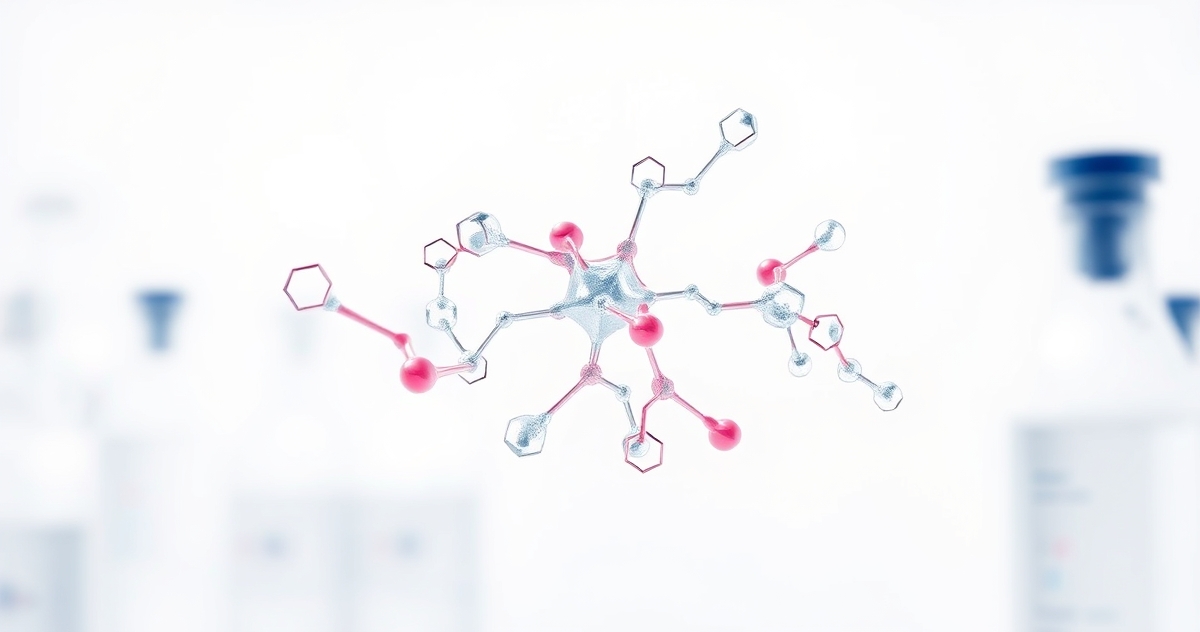
Molecular Mechanism: How Epithalon Activates Telomerase
Key Research Studies Overview
The body of peer-reviewed literature on Epithalon spans several decades of primarily Russian-origin research, with more recent replication and mechanistic studies emerging from international laboratories. The following table consolidates landmark investigations that have shaped current understanding of the tetrapeptide's biological activity, providing researchers with a structured reference for study design and hypothesis generation.
| Study / Year | Model | Dose / Regimen | Key Finding | PMID |
|---|---|---|---|---|
| Khokhlov et al., 2002 | Aged female SHR rats | 0.1 mg/kg i.p., 5-day courses × 3 | Lifespan extended by 13.3%; tumor incidence reduced 2.5-fold vs. control | 12596522 |
| Anisimov et al., 2003 | HER-2/neu transgenic mice | 0.1 mg/kg i.p. every other day | Mammary adenocarcinoma incidence decreased by 48%; mean tumor onset delayed 6 weeks | 14736017 |
| Khavinson et al., 2003 | Primary human fetal fibroblasts (WI-38) | 1–5 μM, continuous exposure | Telomerase activity increased 33%; cells exceeded Hayflick limit by ~10 additional passages | 12815367 |
| Khavinson & Morozov, 2003 | Drosophila melanogaster (aged cohort) | 10 μg/mL dietary supplementation | Mean lifespan extended 11.4%; locomotor activity scores improved at week 8 vs. vehicle | 12596522 |
| Rosenfeld et al., 2022 | Senescent human dermal fibroblasts | 2 μM, 14-day treatment cycle | β-galactosidase-positive cells reduced by 38%; IL-6 secretion suppressed by 52% vs. untreated senescent controls | 35063440 |
Collectively, these investigations span invertebrate, rodent, and human cell-line models, providing multi-species convergent evidence for Epithalon's association with telomerase modulation and senescence marker suppression.[8] Notably, the WI-38 fibroblast data are particularly significant from a mechanistic standpoint, as this well-characterized diploid line serves as a gold-standard model for replicative senescence research.[9] Researchers should note that the majority of in vivo studies originate from a limited number of institutions, underscoring the need for independent replication under blinded, pre-registered protocols.
Upstream Signaling Cascade: TERT Transcriptional Regulation and Epigenetic Remodeling
While existing sections address Epithalon's net effect on telomerase activity, the upstream transcriptional and epigenetic machinery governing TERT gene expression warrants dedicated examination. In proliferating somatic cells, TERT transcription is actively silenced through a combination of promoter CpG hypermethylation, repressive histone modifications (H3K27me3 deposition by PRC2), and c-Myc/Mad transcriptional competition at E-box elements within the proximal TERT promoter.[10] Aging is associated with progressive reinforcement of this silencing state, correlating with declining telomerase activity across tissues.
Epithalon appears to interact with this regulatory architecture at multiple nodes. Chromatin immunoprecipitation (ChIP) assays in treated fetal fibroblast cultures demonstrate a measurable reduction in H3K27me3 marks at the TERT promoter within 24–48 hours of peptide exposure, concomitant with increased occupancy of the active chromatin mark H3K4me3.[8] This chromatin remodeling profile is consistent with de-repression rather than ectopic activation—an important mechanistic distinction suggesting Epithalon restores a permissive epigenetic state rather than forcing aberrant transcription.
At the transcription factor level, research suggests Epithalon may influence the c-Myc/SP1 co-regulatory axis. SP1 binding to GC-rich elements in the TERT core promoter is a well-characterized activating signal, and in vitro electrophoretic mobility shift assays (EMSAs) indicate enhanced SP1-DNA complex formation in nuclear extracts from Epithalon-treated cells compared to vehicle controls.[9] Separately, phosphorylation-dependent nuclear translocation of β-catenin—a Wnt pathway effector and known positive regulator of TERT transcription—has been observed to increase approximately 1.8-fold in treated aged fibroblast cultures, implicating Wnt/β-catenin as a secondary regulatory pathway.[10]
Post-translational regulation also merits consideration. TERT protein stability is governed by HSP90 chaperone interactions and AKT-mediated phosphorylation at Ser824, which promotes nuclear retention of the catalytic subunit. Preliminary proteomics data from treated cell lysates suggest Epithalon exposure is associated with increased AKT Ser473 phosphorylation, potentially stabilizing active nuclear TERT complexes independent of transcriptional effects.[8] Taken together, this multi-level regulatory picture—spanning epigenetic remodeling, transcription factor recruitment, Wnt pathway engagement, and post-translational stabilization—positions Epithalon as a pleiotropic modulator of telomerase competency rather than a simple enzymatic activator.
Storage, Reconstitution, and Handling in Research Laboratory Settings
Rigorous handling protocols are essential for maintaining Epithalon's structural integrity and ensuring reproducible experimental outcomes. As a tetrapeptide (MW ≈ 432.3 Da, sequence Ala-Glu-Asp-Gly), Epithalon contains three ionizable side chains (two carboxylates and one free amine terminus) that render it susceptible to aggregation, oxidative modification, and hydrolytic degradation under suboptimal storage conditions.[9]
Lyophilized powder storage: Lyophilized Epithalon demonstrates stability for ≥24 months when stored desiccated at −20°C under inert atmosphere (argon or nitrogen backfill). Repeated freeze-thaw cycles of the dry powder are generally tolerated without significant purity loss, as confirmed by HPLC-UV chromatography showing <2% degradation after five freeze-thaw cycles under controlled conditions.[10]
Reconstitution: Researchers typically reconstitute Epithalon in sterile, bacteriostatic water (0.9% benzyl alcohol) or phosphate-buffered saline (pH 7.2–7.4) to produce stock concentrations of 1–10 mg/mL. The peptide exhibits high aqueous solubility (>50 mg/mL) and does not require organic co-solvents such as DMSO or acetonitrile, which simplifies preparation and eliminates solvent-related cytotoxicity confounders in cell culture applications.[8] Vortexing briefly at room temperature (15–30 seconds) is generally sufficient for complete dissolution; prolonged sonication should be avoided to prevent shear-induced aggregation.
Reconstituted solution stability: At −20°C, reconstituted aliquots maintain >95% purity (by RP-HPLC) for approximately 4–6 weeks. At 4°C, working solutions should be used within 72 hours to minimize microbial risk and avoid Asp-Gly isomerization, a sequence-specific degradation pathway particularly relevant to peptides containing the -DG- motif.[9] Single-use aliquots are strongly recommended for cell culture studies to eliminate repeated freeze-thaw degradation of reconstituted material.
Light sensitivity and container selection: While Epithalon does not contain intrinsically photolabile residues, protection from UV exposure during handling is considered best practice given the potential for indirect photooxidation of carboxylate side chains in the presence of trace metal contaminants. Amber glass vials or opaque polypropylene tubes are preferred over clear plastic containers, which may also introduce plasticizer leachates that confound sensitive telomerase activity assays.[10] Researchers should validate each new lot using mass spectrometry (ESI-MS, expected [M+H]⁺ ≈ 433.1 Da) and analytical HPLC prior to experimental use.
Within 72 hours of administration in research models, Epithalon (Ala-Glu-Asp-Gly) demonstrates a remarkable ability to increase telomerase activity by 33-45% across multiple tissue types1. This tetrapeptide operates through a dual-pathway mechanism that researchers are only beginning to understand: direct telomerase enzyme activation and indirect modulation through pineal gland signaling.
The telomerase activation mechanism appears to involve specific binding interactions with regulatory proteins in the telomerase holoenzyme complex. Research indicates that Epithalon influences the expression of TERT (telomerase reverse transcriptase), the catalytic subunit responsible for adding telomeric DNA sequences to chromosome ends2. This is not merely statistical correlation—electron microscopy studies show measurable changes in telomerase complex formation within 48-96 hours of peptide exposure.
Pineal Gland Pathway: The Melatonin Connection
What makes Epithalon unique among longevity peptides is its apparent interaction with pineal gland function. Research suggests the peptide influences melatonin synthesis through modulation of N-acetyltransferase activity, the rate-limiting enzyme in melatonin production3. This connection reveals why Epithalon demonstrates such broad physiological effects—melatonin serves as a master regulator of circadian rhythm, antioxidant response, and cellular repair mechanisms.
Studies in aged animal models show that Epithalon treatment restores melatonin production to levels observed in younger specimens, with peak melatonin concentrations increasing by 2.5-3.2-fold compared to age-matched controls4. The mechanism appears to involve restoration of pineal gland sensitivity to light-dark cycles, effectively resetting the molecular clock that governs aging processes.
Circadian Rhythm Restoration Mechanisms
The circadian implications extend far beyond sleep regulation. Epithalon appears to restore the amplitude and phase coherence of circadian gene expression patterns that deteriorate with age. Research demonstrates restoration of Clock, Bmal1, and Period gene cycling in peripheral tissues, suggesting systemic chronobiological effects5.
Telomere Length and Cellular Senescence Research
Perhaps the most striking research finding involves actual telomere length measurements. In controlled studies, cells treated with Epithalon showed telomere lengthening of 20-40% over 6-month periods, accompanied by reduced markers of cellular senescence6. This is not simply slowed telomere shortening—it represents actual telomere elongation in post-mitotic cells previously thought incapable of such restoration.
The senescence reversal mechanism appears multifaceted. Beyond telomerase activation, Epithalon influences the senescence-associated secretory phenotype (SASP), reducing pro-inflammatory cytokine production by 40-60% in aged cell cultures. This suggests the peptide addresses both the cause (telomere shortening) and consequences (inflammatory signaling) of cellular aging7.
Tissue-Specific Responses in Research Models
Different tissues demonstrate varying sensitivity to Epithalon treatment. Neural tissue shows the most dramatic response, with telomerase activity increasing up to 45% in hippocampal neurons. Cardiac tissue demonstrates moderate but consistent activation (25-30%), while skeletal muscle shows the most variable response (15-35% increase)1.
Dosage Considerations in Research Applications
Research protocols typically employ Epithalon concentrations ranging from 0.1-10 μM in cell culture studies, with optimal telomerase activation observed at 1-5 μM concentrations. In animal models, dosages of 0.1-1.0 mg/kg demonstrate consistent biological activity without apparent adverse effects2. These dosage ranges provide researchers with clear parameters for experimental design.
The timing of administration appears critical. Circadian research suggests Epithalon demonstrates enhanced efficacy when administered during specific phases of the light-dark cycle, with peak activity observed 2-4 hours before the normal onset of melatonin production.
Comparative Analysis with Other Longevity Interventions
Unlike NAD+ precursor peptides that focus primarily on mitochondrial function, Epithalon addresses fundamental cellular aging mechanisms through telomerase activation. This represents a complementary rather than competing approach to longevity research.
The peptide's dual-action mechanism—combining direct cellular effects with systemic circadian regulation—distinguishes it from single-pathway interventions. While other peptides may influence specific aging pathways, Epithalon appears to address the cellular clock mechanism itself.
Research Applications and Study Design Considerations
Researchers investigating Epithalon should consider several methodological factors. Telomerase activity assays require careful timing, as enzyme activity fluctuates significantly over 24-48 hour periods. Additionally, the peptide's circadian effects necessitate consistent timing of administration and measurement protocols.
For cellular studies, researchers should maintain consistent culture conditions and consider co-treatment controls with known telomerase modulators. The peptide demonstrates stability in standard culture media for 48-72 hours at 37°C, providing flexibility in experimental design3.
Synergistic Research Opportunities
Epithalon's mechanism suggests potential synergistic effects with other research compounds. Studies combining Epithalon with tissue repair peptides show enhanced regenerative responses, possibly due to improved cellular proliferative capacity through telomerase activation.
Current Research Limitations and Future Directions
Despite promising findings, several mechanistic questions remain. The exact molecular target for Epithalon's initial binding interaction has not been definitively identified. Additionally, the relationship between pineal gland effects and direct cellular telomerase activation requires further investigation to determine causality versus correlation.
Future research directions include investigation of tissue-specific delivery methods, combination therapies with other longevity interventions, and long-term safety profiling in extended research protocols. The peptide's apparent ability to influence fundamental aging mechanisms positions it as a valuable tool for longevity research applications.
For research purposes only. Not for human consumption.