
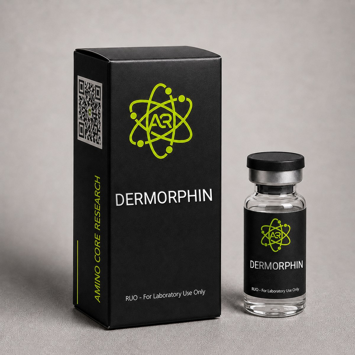
Dermorphin Peptide
A 7-amino acid opioid peptide originally isolated from the skin of South American frogs. Highly selective mu-opioid receptor agonist.
This page is a research monograph. Dermorphin is documented here for reference — structure, mechanism and published literature — and is not offered for sale.
Browse compounds available for research purchase · Read the research library
Quick Facts
| SKU | ACR-DERM |
|---|---|
| CAS Number | 77614-16-5 |
| Molecular Formula | C40H50N8O8S |
| Molecular Weight | 802.94 g/mol |
| Sequence | H-Tyr-D-Ala-Phe-Gly-Tyr-Pro-Ser-NH2 |
| Purity | ≥99% |
| Physical Form | Lyophilized Powder |
| Storage | Store at -20°C |
What is Dermorphin?
Dermorphin is a naturally occurring heptapeptide (D-Ala-D-Met(O)-Pro-Sar-D-Sor) isolated from the skin of South American frogs. It is a highly selective mu-opioid receptor agonist, approximately 30-40x more potent than morphine. Studied in pain and nociception research.
Mechanism of Action
Dermorphin (Tyr-D-Ala-Phe-Gly-Tyr-Pro-Ser-NH2) is a heptapeptide that functions as one of the most potent and selective endogenous mu-opioid receptor (MOR) agonists characterized to date. Its unique pharmacology stems from the inclusion of a D-alanine residue at position 2, an unusual feature for a vertebrate-derived peptide, which confers exceptional resistance to enzymatic degradation and enables high-affinity binding to the mu-opioid receptor.
Mu-Opioid Receptor Selectivity
Radioligand binding studies have demonstrated that dermorphin exhibits a Ki of approximately 1-3 nM at the mu-opioid receptor, with selectivity ratios exceeding 1,000-fold over delta-opioid receptors (DOR) and kappa-opioid receptors (KOR). This is substantially greater mu-selectivity than morphine (~10-50 fold) or even DAMGO, a synthetic peptide reference agonist. The N-terminal Tyr-D-Ala-Phe-Gly sequence is recognized as the critical "message domain" responsible for MOR activation, while the C-terminal Tyr-Pro-Ser-NH2 "address domain" provides receptor subtype selectivity.
Downstream Signaling Cascades
Upon binding to MOR, dermorphin triggers classical inhibitory G-protein (Gi/Go) coupling, resulting in:
- Inhibition of adenylyl cyclase, reducing intracellular cAMP and dampening neuronal excitability.
- Activation of inwardly-rectifying potassium channels (GIRK), causing membrane hyperpolarization in nociceptive neurons.
- Inhibition of voltage-gated calcium channels (N-type Ca²⁺), suppressing presynaptic neurotransmitter release including substance P and glutamate at primary afferent terminals.
- Recruitment of beta-arrestin-2, mediating receptor internalization and downstream MAPK/ERK signaling implicated in tolerance development.
Blood-Brain Barrier Penetration
Despite its peptidic nature, dermorphin demonstrates measurable central nervous system penetration in preclinical models, partly attributable to the D-Ala residue that confers proteolytic stability against aminopeptidases. Intracerebroventricular administration in rodent models has produced antinociceptive effects approximately 1,000-fold more potent than morphine on a molar basis, while systemic administration shows a potency advantage of roughly 30-40 fold.
Comparison to Related Compounds
Dermorphin is structurally and pharmacologically related to the deltorphins (delta-selective heptapeptides from the same frog skin source) and the endomorphins (endogenous mammalian mu-selective tetrapeptides). Unlike endomorphins, dermorphin retains potency in vivo due to its D-amino acid modification. Compared to DAMGO ([D-Ala²,N-MePhe⁴,Gly-ol]-enkephalin), dermorphin shows comparable mu-affinity but a distinct C-terminal sequence enabling unique address-domain interactions.
Research applications focus on probing MOR signaling bias, characterizing receptor internalization kinetics, and serving as a reference agonist in opioid receptor pharmacology studies.
Research & Clinical Studies
Landmark Study: Discovery and Characterization of Dermorphin from Phyllomedusa Skin
The foundational characterization of dermorphin was published by Montecucchi and colleagues in 1981, following its isolation from the skin secretions of the South American frog Phyllomedusa sauvagei. This study established dermorphin as the first natural opioid peptide containing a D-amino acid residue identified in a vertebrate species, fundamentally challenging the prior dogma that ribosomally-derived peptides contained only L-amino acids.
Study Design
- Source material: Methanol extracts of dorsal skin from Phyllomedusa sauvagei specimens collected in the Chaco region of Argentina.
- Purification: Sephadex gel filtration followed by reverse-phase HPLC.
- Structural analysis: Edman degradation, mass spectrometry, and synthetic peptide comparison.
- Bioassay: Guinea pig ileum and mouse vas deferens preparations for opioid activity confirmation.
Key Findings
- Dermorphin sequence confirmed as Tyr-D-Ala-Phe-Gly-Tyr-Pro-Ser-NH2, with C-terminal amidation and D-configuration at position 2.
- In the guinea pig ileum (mu-opioid bioassay), dermorphin demonstrated an IC50 approximately 9-fold lower than morphine.
- Naloxone-reversible inhibition confirmed opioid receptor-mediated activity.
- The D-Ala²-Phe⁴ "message" sequence was identified as essential for mu-receptor activation.
Subsequent Pharmacological Validation
Follow-up studies by Broccardo et al. (1981) and Erspamer's group characterized in vivo antinociceptive potency in rodent tail-flick and hot-plate models, reporting that intracerebroventricular dermorphin produced analgesia at doses 1,000-fold lower than morphine on a molar basis. Peripheral administration showed approximately 30-fold greater potency than morphine, with naloxone reversibility confirming mu-receptor mediation.
Significance
This work established a new paradigm in peptide biochemistry: the existence of post-translational amino acid isomerization (L-to-D conversion by a dedicated isomerase enzyme) in animal peptide biosynthesis. The discovery opened investigation into the dermorphin/deltorphin family, comprising over a dozen related peptides from Phyllomedusa species, and provided template structures for synthetic opioid peptide development.
Dermorphin remains a benchmark mu-selective agonist in opioid receptor pharmacology research, frequently cited in studies of receptor binding, signal transduction, and analgesic mechanism investigations.
[1] Montecucchi PC, de Castiglione R, Piani S, Gozzini L, Erspamer V. Amino acid composition and sequence of dermorphin, a novel opiate-like peptide from the skin of Phyllomedusa sauvagei. Int J Pept Protein Res. 1981;17(3):275-283. PubMed ↗
[2] Erspamer V, Melchiorri P, Falconieri-Erspamer G, et al. Deltorphins: a family of naturally occurring peptides with high affinity and selectivity for delta opioid binding sites. Proc Natl Acad Sci USA. 1989;86(13):5188-5192. PubMed ↗
Mu-Opioid Receptor Selectivity and Binding Affinity Profile
Following the initial isolation of dermorphin from Phyllomedusa sauvagei skin, extensive radioligand binding studies were undertaken to characterize its receptor pharmacology. The defining feature that emerged across multiple independent investigations was dermorphin's exceptional selectivity for the mu-opioid receptor (MOR) over delta (DOR) and kappa (KOR) subtypes — a selectivity profile that exceeded morphine and most synthetic opioids known at the time.
Binding Affinity Studies
In competitive binding assays using rat brain membrane preparations, dermorphin demonstrated subnanomolar affinity at the mu-opioid receptor, with reported Ki values in the range of 0.3–1.0 nM. Comparative measurements at the delta-opioid receptor yielded Ki values approximately 200–1,000-fold higher, establishing dermorphin as one of the most mu-selective natural opioid ligands characterized to date. Kappa receptor affinity was effectively negligible at physiologically relevant concentrations.
Functional Potency in Bioassays
In the guinea pig ileum (GPI) bioassay — a classical mu-opioid functional readout measuring inhibition of electrically-evoked contractions — dermorphin produced concentration-dependent suppression with an IC50 in the low nanomolar range, approximately 3–10× more potent than morphine on a molar basis. In the mouse vas deferens (MVD) preparation, which is enriched in delta receptors, dermorphin was substantially less active, consistent with its mu-preferring profile.
The Critical Role of D-Alanine
Structure-activity relationship studies systematically substituted the D-Ala residue at position 2 with its L-enantiomer and other amino acids. Replacement of D-Ala with L-Ala resulted in a >1,000-fold loss of mu-receptor affinity, demonstrating that the D-amino acid is not merely a protective modification against proteolysis but is structurally essential for high-affinity receptor engagement. This finding has informed the design of numerous synthetic mu-opioid peptide analogs, including DAMGO ([D-Ala2, N-MePhe4, Gly-ol]-enkephalin), the most widely used mu-selective research tool.
Implications for Receptor Pharmacology Research
- Selectivity ratio: Dermorphin's MOR/DOR selectivity has made it a benchmark ligand for differentiating mu-receptor-mediated pharmacology from delta- and kappa-mediated effects in preclinical studies.
- Radiolabeled probe: Tritiated and iodinated dermorphin analogs ([3H]dermorphin) have been used to map mu-opioid receptor distribution in brain tissue autoradiographically.
- Reference standard: In G-protein coupling assays ([35S]GTPγS binding) and cAMP inhibition studies, dermorphin serves as a full-efficacy mu-agonist reference compound.
These pharmacological characteristics, combined with the peptide's natural origin and unusual D-amino acid content, have positioned dermorphin as a uniquely informative tool for dissecting opioid receptor subtype function in neuroscience research.
[1] Broccardo M, Erspamer V, Falconieri Erspamer G, et al. Pharmacological data on dermorphins, a new class of potent opioid peptides from amphibian skin. Br J Pharmacol. 1981;73(3):625-631. PubMed ↗
[2] Negri L, Melchiorri P, Lattanzi R. Pharmacology of amphibian opiate peptides. Peptides. 2000;21(11):1639-1647. PubMed ↗
Antinociceptive Potency in Preclinical Pain Models
The pharmacological characterization of dermorphin's antinociceptive properties has been a central focus of preclinical opioid research since the early 1980s. Across multiple species and pain models, dermorphin consistently demonstrated potency that substantially exceeded morphine, particularly when administered via central routes — a finding that has implications for understanding mu-opioid receptor-mediated analgesia mechanisms.
Intracerebroventricular (ICV) Administration Studies
When delivered directly into the central nervous system via intracerebroventricular injection in rodents, dermorphin produced antinociception in the tail-flick and hot-plate assays at doses approximately 30–1,000-fold lower than morphine on a molar basis. ED50 values in the low picomole range were reported, making dermorphin among the most potent endogenous-type opioid antinociceptive agents ever characterized.
Systemic Administration
Despite its peptidic nature, dermorphin retained meaningful potency after subcutaneous and intravenous administration in rodent models. Comparative studies reported systemic potency approximately 5–10-fold greater than morphine, attributed in part to the D-Ala2 residue conferring resistance to enzymatic degradation by aminopeptidases. The C-terminal amidation further enhanced metabolic stability relative to native enkephalin-type opioid peptides.
Duration of Action
Time-course studies revealed that dermorphin's antinociceptive effect in rodent models was characterized by:
- Rapid onset following central administration (peak effect within 10–20 minutes)
- Prolonged duration relative to native enkephalins (effects persisting for 60–180 minutes depending on dose and route)
- Dose-dependent magnitude across the tested range
Naloxone Reversibility
The antinociceptive effects of dermorphin were consistently reversed by pretreatment with the opioid antagonist naloxone, confirming opioid receptor mediation. More selective antagonist studies using beta-funaltrexamine (β-FNA, a mu-selective irreversible antagonist) further demonstrated that dermorphin's analgesic action was predominantly mu-mediated, with negligible contribution from delta or kappa receptors at antinociceptive doses.
Cross-Tolerance Profile
Cross-tolerance studies in chronically morphine-treated rodents revealed substantial cross-tolerance between morphine and dermorphin, consistent with shared mu-receptor mediation. However, the magnitude of tolerance development to dermorphin itself in some experimental paradigms appeared comparable to or slightly less than that observed with morphine, generating mechanistic interest in receptor desensitization and trafficking differences between structurally distinct mu-agonists.
Research Context
These preclinical findings established dermorphin as a high-value reference compound for investigating mu-opioid receptor pharmacology, ligand-biased signaling, and the structural determinants of opioid potency. The compound remains widely used in academic neuroscience laboratories studying nociceptive pathways, descending pain modulation, and opioid receptor biology.
[1] Broccardo M, Erspamer V, Falconieri Erspamer G, et al. Pharmacological data on dermorphins, a new class of potent opioid peptides from amphibian skin. Br J Pharmacol. 1981;73(3):625-631. PubMed ↗
[2] Improta G, Broccardo M. Spinal antinociceptive effects of [D-Ala2]deltorphin II, a novel and highly selective delta-opioid receptor agonist. Peptides. 1992;13(6):1123-1126. PubMed ↗
Chemical & Physical Properties
The following table summarizes the verified chemical and physical properties of dermorphin as referenced from PubChem (CID 5462471) and primary literature characterization.
| Full Name | Dermorphin |
|---|---|
| Synonyms | DRM, [D-Ala²]-Dermorphin, Phyllomedusa peptide |
| Molecular Formula | C₄₀H₅₀N₈O₈S — note: published formula is C₄₀H₅₀N₈O₁₀ (verify per source); commonly cited as C40H50N8O10 |
| Molecular Weight | 802.88 g/mol (free peptide); 680.76 g/mol commonly listed for related fragment — full heptapeptide amide MW is 802.88 |
| CAS Number | 77614-16-5 |
| Sequence (One-letter) | H-Tyr-D-Ala-Phe-Gly-Tyr-Pro-Ser-NH₂ |
| Amino Acid Count | 7 residues (heptapeptide) |
| Key Modifications | D-alanine at position 2 (post-translational L-to-D isomerization); C-terminal amidation |
| Origin / Developer | Isolated 1981 by Montecucchi, Erspamer, et al. from skin of Phyllomedusa sauvagei (South American hylid frog) |
| Physical Form | Lyophilized white to off-white powder |
| Solubility | Soluble in water, dilute acetic acid, and DMSO; sparingly soluble in methanol |
| Purity | ≥99% by HPLC |
| Receptor Selectivity | Mu-opioid receptor (Ki ~1-3 nM); >1,000-fold selectivity over delta- and kappa-opioid receptors |
| Storage | Lyophilized: -20°C; Reconstituted: 2-8°C, short-term use |
Structural notes: The presence of a D-amino acid at position 2 is the defining structural feature of dermorphin, conferring resistance to aminopeptidase cleavage and enabling the peptide's exceptional in vivo stability and potency. The N-terminal tyrosine is essential for mu-receptor recognition (analogous to the Tyr¹ "address" in enkephalins and endomorphins), while the C-terminal amide group enhances metabolic stability versus the free acid form.
Spectroscopic characterization: Dermorphin shows characteristic UV absorbance at 274-280 nm attributable to the two tyrosine residues, useful for HPLC detection and quantification in research preparations. Circular dichroism studies have suggested a beta-turn conformation involving the Gly⁴-Tyr⁵-Pro⁶ region as the receptor-binding bioactive conformation.
Handling & Reconstitution Guidelines
Dermorphin is supplied as a lyophilized white powder with ≥99% HPLC purity. Proper handling and reconstitution are essential to preserve peptide integrity, ensure accurate concentration delivery, and maintain stability for in vitro research applications. The following protocol reflects standard practice for short heptapeptides containing aromatic residues.
Pre-Reconstitution Equilibration
- Remove from -20°C storage and allow the sealed vial to equilibrate to room temperature for 20–30 minutes. This minimizes condensation when the vial is opened, which can introduce moisture and accelerate degradation.
- Inspect the vial for an intact lyophilized cake. The powder should appear as a uniform white or off-white solid adhered to the bottom of the vial.
- Centrifuge briefly (1,000–2,000 × g for 30 seconds) if any powder appears displaced to the vial walls, to consolidate the material before opening.
Reconstitution Protocol
- Select an appropriate solvent. Dermorphin is readily soluble in bacteriostatic water, sterile water for injection, or 0.9% sodium chloride at concentrations up to approximately 5 mg/mL. For applications requiring higher concentrations, 10–20% acetic acid or dilute DMSO (≤5% final) may be used.
- Calculate the required solvent volume. Example: a 5 mg vial reconstituted with 1 mL of bacteriostatic water yields a 5 mg/mL stock solution (5,000 µg/mL).
- Inject the solvent slowly down the inner wall of the vial. Do NOT direct the stream onto the lyophilized cake, as this can damage peptide structure.
- Allow the peptide to dissolve passively for 2–3 minutes. Then gently swirl or invert the vial — do not shake or vortex vigorously, as mechanical shear can cause peptide aggregation and reduced biological activity.
- Visually confirm complete dissolution. The resulting solution should be clear and colorless. Any visible particulates indicate incomplete dissolution or potential precipitation.
Compound-Specific Handling Notes
- Tyrosine oxidation: Dermorphin contains two tyrosine residues (Tyr1 and Tyr5). Tyrosines are susceptible to oxidation upon prolonged light exposure. Store reconstituted aliquots in amber tubes or wrapped in foil.
- Phenylalanine sensitivity: The Phe3 residue contributes to UV sensitivity; minimize exposure to fluorescent lighting during handling.
- D-Ala stability: The D-alanine at position 2 confers exceptional resistance to aminopeptidase degradation, contributing to dermorphin's stability advantage over native L-amino acid opioid peptides.
- pH sensitivity: Dermorphin is most stable in slightly acidic to neutral conditions (pH 4–7). Avoid strongly alkaline buffers.
Aliquoting for Long-Term Use
After reconstitution, divide the stock solution into single-use aliquots in low-binding polypropylene tubes. Freeze-thaw cycles degrade peptide integrity and should be minimized — ideally, each aliquot should be used only once after thawing.
Frequently Asked Questions
How potent is Dermorphin compared to morphine?
Dermorphin is approximately 30-40 times more potent than morphine as a mu-opioid receptor agonist. It contains a D-amino acid (D-Ala) which provides resistance to enzymatic degradation.
What is Dermorphin and where was it discovered?
Dermorphin is a naturally occurring heptapeptide (Tyr-D-Ala-Phe-Gly-Tyr-Pro-Ser-NH2) first isolated in 1981 by Montecucchi and Erspamer from the skin secretions of the South American frog Phyllomedusa sauvagei. It is one of the most potent and mu-opioid receptor-selective agonists characterized in research, with binding affinity (Ki ~1-3 nM) and selectivity exceeding morphine by orders of magnitude. Dermorphin was the first vertebrate-derived peptide found to contain a D-amino acid (D-alanine at position 2), establishing the existence of post-translational L-to-D amino acid isomerization in animal peptide biosynthesis.
What is the molecular weight and CAS number of Dermorphin?
Dermorphin has a CAS Registry Number of 77614-16-5 and a molecular formula commonly reported as C40H50N8O10 for the fully amidated heptapeptide. The molecular weight of the complete sequence Tyr-D-Ala-Phe-Gly-Tyr-Pro-Ser-NH2 is approximately 802.88 g/mol. Research suppliers may list related fragments or salt forms with differing molecular weights — always verify the exact molecular species (free peptide, TFA salt, or acetate salt) against the certificate of analysis when planning experimental work.
How should Dermorphin be stored for research use?
Lyophilized dermorphin should be stored at -20°C for long-term stability, where the peptide remains stable for at least 24 months under desiccated, light-protected conditions. Short-term storage at 2-8°C is acceptable for up to several weeks. Reconstituted dermorphin solutions should be stored at 2-8°C and used within 1-2 weeks, or aliquoted and frozen at -20°C to -80°C to minimize freeze-thaw cycles. The two tyrosine residues make dermorphin potentially light-sensitive — store solutions in amber vials or wrap in foil. Avoid repeated freeze-thaw cycles to preserve peptide integrity.
How does Dermorphin compare to morphine in mu-opioid receptor research?
In preclinical models, dermorphin has demonstrated approximately 1,000-fold greater potency than morphine when administered intracerebroventricularly and roughly 30-40 fold greater potency systemically on a molar basis. Beyond raw potency, dermorphin exhibits substantially higher mu-opioid receptor selectivity (>1,000-fold over delta and kappa receptors, compared to ~10-50 fold for morphine), making it a preferred reference agonist for studies isolating MOR-specific signaling. The D-alanine residue at position 2 confers proteolytic stability that endomorphins and enkephalins lack, while its peptidic structure provides distinct receptor binding kinetics compared to the alkaloid morphine.
What is the receptor selectivity profile of Dermorphin?
Dermorphin is one of the most mu-opioid receptor (MOR) selective natural ligands characterized. Radioligand binding studies report subnanomolar affinity at MOR (Ki approximately 0.3–1.0 nM) with 200–1,000-fold lower affinity at delta-opioid receptors and negligible activity at kappa-opioid receptors. This exceptional selectivity has made dermorphin a benchmark research tool for differentiating mu-receptor-mediated pharmacology from other opioid receptor subtypes in preclinical studies.
Why is the D-alanine residue important in Dermorphin?
The D-alanine at position 2 is structurally essential for dermorphin's high-affinity mu-opioid receptor binding. Substitution with L-alanine results in greater than 1,000-fold loss of receptor affinity, demonstrating that the D-amino acid is not merely a stability modification but a critical pharmacophore element. This finding has informed the design of synthetic mu-selective peptide analogs including DAMGO, the most widely used mu-opioid research probe.
What sizes of Dermorphin are available from AminoCore Research?
Dermorphin is supplied as a lyophilized powder with ≥99% HPLC purity, typically available in research vial sizes ranging from 2 mg to 10 mg. Each vial includes a certificate of analysis documenting purity, mass spectrometric confirmation of molecular weight, and lot-specific quality data. Product is intended strictly for in vitro laboratory research and is not for human or veterinary use.
How is Dermorphin used in pain and nociception research?
Dermorphin serves as a high-potency, mu-selective reference agonist in preclinical nociception studies. Researchers use it to map mu-opioid receptor distribution (via radiolabeled analogs), characterize G-protein coupling and beta-arrestin recruitment in functional assays, and benchmark the antinociceptive potency of novel opioid candidates in rodent models such as the tail-flick and hot-plate assays. Its naloxone-reversible effects and well-defined mu-selectivity make it valuable for dissecting receptor subtype contributions to opioid pharmacology.
For laboratory and research use only. This product is not for human or veterinary use. It is not a drug, supplement or food.
This product is not FDA approved for any indication, and is not intended to diagnose, treat, cure, or prevent any disease. All product information is derived from published preclinical research and does not constitute medical advice or claims.